Categories Slip Disc/Sciatica Pain

Slip Disc Recovery Time: What to Expect Week by Week

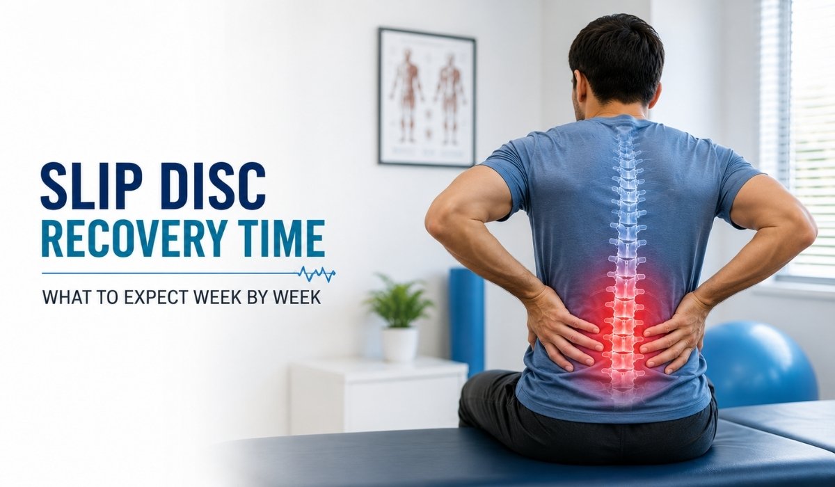
Week-by-week recovery timeline for slip disc treatment showing pain relief, healing stages, and return to daily activities

Quick Answer

Most people with a slip disc (herniated or prolapsed disc) notice meaningful pain relief within 4–6 weeks of starting conservative treatment such as physiotherapy, activity modification and, if needed, targeted injections. About 80–90% of patients improve without surgery within 3–6 months. Recovery speed depends on the disc level involved, the size of the herniation, and how consistently the treatment plan is followed.

Quick Summary

  • Bed rest is not the goal — controlled movement from week one usually speeds recovery.
  • Most patients see noticeable improvement within 4–6 weeks of starting treatment.
  • Sciatica-type leg pain typically takes longer to settle than localised back pain — often 6–12 weeks.
  • Structured physiotherapy usually begins in the first 1–2 weeks, not after pain disappears.
  • Full return to physically demanding work or sport can take 3–6 months.
  • Numbness, progressive weakness, or bladder/bowel changes need same-day medical evaluation.
  • Recovery timelines are individual — age, disc level, general fitness, and treatment adherence all matter.
  • A pain specialist can tailor the plan and adjust it if progress stalls at any stage.

What Is a Slip Disc?

A “slip disc” (medically called a herniated, prolapsed, or bulging disc) occurs when the soft inner core of a spinal disc pushes through a weakened outer layer. This can irritate nearby nerves, most often in the lower back (lumbar spine), causing localised pain, stiffness, and sometimes pain that radiates into the leg (sciatica). The disc doesn’t literally “slip” out of place — the term describes the bulge or rupture of disc material.

Symptoms to Watch For

  • Localised lower back or neck pain that may worsen with sitting or bending
  • Radiating pain into the buttock, leg, or arm depending on the disc level
  • Tingling, numbness, or a burning sensation along the nerve pathway
  • Muscle weakness in the leg or foot in more significant cases
  • Pain that eases with lying down and worsens with prolonged standing or coughing

Causes and Risk Factors

Disc herniation is usually the result of gradual wear (disc degeneration) combined with a triggering movement, rather than a single dramatic injury. Common contributors include:

  • Age-related disc dehydration and reduced flexibility (most common after 30–50 years)
  • Repetitive bending, twisting, or heavy lifting with poor technique
  • Prolonged sitting with poor posture, common in desk-based jobs
  • Excess body weight, which increases load on lumbar discs
  • Smoking, which reduces disc nutrition and healing capacity
  • Sedentary lifestyle with weak core and back-supporting muscles

How a Slip Disc Is Diagnosed

Diagnosis starts with a clinical history and physical examination, including tests for nerve involvement such as leg-raise tests and reflex checks. Imaging — typically an MRI — is used to confirm the disc level, the size of the herniation, and whether a nerve is being compressed. Nexus Pain does not recommend imaging or invasive treatment for every mild case; many patients recover with structured conservative care alone, and imaging is reserved for those with red-flag symptoms or pain that doesn’t respond to initial treatment.

Slip Disc Recovery Time: Week-by-Week

The exact numbers vary by patient, but this is the general pattern most people can expect once appropriate treatment begins.

Week 1–2: Acute Phase — Pain Control and Gentle Movement

The priority is calming inflammation and avoiding positions that aggravate the nerve, while staying gently active. Complete bed rest beyond a day or two is generally discouraged, as prolonged inactivity can slow recovery and weaken supporting muscles. Short, frequent walks, anti-inflammatory measures, and activity modification are typical. A physiotherapy assessment is often started during this window.

Week 3–4: Early Rehabilitation

Pain usually starts to shift from constant to intermittent, and leg or arm symptoms may begin to reduce as inflammation settles. Guided physiotherapy — nerve-gliding exercises, gentle stretching, and posture correction — typically progresses here. Patients who skip this phase or return to full activity too soon are more likely to see symptoms flare up again.

Week 5–8: Functional Recovery

Most patients notice a clear improvement in day-to-day comfort by this stage, with reduced reliance on medication. Core-strengthening and postural correction exercises are progressed, and light work duties can often resume with ergonomic adjustments. Persistent or worsening leg pain at this stage is a signal to reassess the treatment plan rather than “wait it out.”

Week 9–12: Consolidation

By around three months, the majority of patients following a structured programme report significant relief and improved function. Return to moderate exercise, longer working hours, and most daily activities is usually appropriate, guided by physiotherapy progress rather than a fixed calendar date.

Months 3–6: Full Functional Return

For physically demanding jobs, contact sports, or heavy lifting, full return typically falls in this window, once strength and flexibility have been rebuilt and confirmed through functional testing. A minority of patients with larger herniations or incomplete relief may be evaluated for further options such as targeted injections or, rarely, surgery, at this stage.

Comparison: Recovery Timeline by Approach

ApproachTypical Timeline to Meaningful ReliefNotes
Activity modification + physiotherapy alone6–12 weeksEffective for the majority of mild-to-moderate cases
Physiotherapy + epidural steroid injection3–6 weeksConsidered when leg/arm pain limits participation in physiotherapy
Radiofrequency ablation (select cases)2–4 weeks post-procedureUsed for specific facet-related or chronic pain patterns, not routine disc herniation
Surgical discectomy (reserved cases)4–6 weeks initial, 3–6 months full recoveryConsidered after failed conservative care or with red-flag symptoms

Non-Surgical Treatment Options

  • Structured physiotherapy: nerve mobilisation, core strengthening, posture retraining
  • Activity modification: avoiding prolonged sitting, heavy lifting, and high-impact loading
  • Medication: short-term anti-inflammatory or nerve-pain medication as prescribed
  • Epidural steroid injections: for significant nerve-related pain limiting rehabilitation
  • Ergonomic and lifestyle adjustments: workstation setup, sleep posture, weight management

Prevention Tips During and After Recovery

  • Maintain a strong core through guided, progressive exercise
  • Use correct lifting technique — bend at the knees, keep the load close to the body
  • Avoid prolonged static sitting; take short movement breaks every 30–45 minutes
  • Address body weight and smoking, both of which affect disc health and healing
  • Continue maintenance exercises after symptoms resolve, not just during flare-ups

Expert Insights

Expert Insight 1: Movement Is Medicine in the Early Phase

Patients often assume rest equals recovery. In reality, controlled movement in the first two weeks helps maintain disc nutrition and prevents stiffness that can prolong recovery. The goal is modified activity, not immobility.


Expert Insight 2: Leg Pain Often Takes Longer Than Back Pain

When a disc herniation irritates a nerve root, the resulting leg or arm pain (sciatica) can lag behind the back pain itself, sometimes taking several extra weeks to settle even as the back feels better. This is a normal pattern, not a sign that treatment has failed.


Expert Insight 3: Consistency Matters More Than Intensity

Patients who complete short, regular physiotherapy sessions tend to progress more predictably than those who do occasional intense sessions. Recovery timelines quoted in research reflect consistent adherence to a structured plan, not sporadic effort.

Common Mistakes That Slow Recovery

  • Prolonged bed rest beyond the first day or two, which weakens supporting muscles
  • Returning to heavy lifting or high-impact exercise as soon as pain reduces, before strength is rebuilt
  • Stopping physiotherapy once acute pain settles, instead of completing the full rehabilitation programme
  • Relying only on painkillers without addressing posture, ergonomics, or core strength

Myths vs. Facts

MythFact
A slip disc always requires surgery.Most cases (roughly 80–90%) improve with non-surgical treatment; surgery is reserved for specific situations.
Complete bed rest heals a slip disc fastest.Extended bed rest can slow recovery; guided, gradual movement is generally more effective.
Once pain disappears, the disc is fully healed.Pain relief often precedes full tissue healing and strength recovery — the rehabilitation programme should still be completed.
A slip disc means you can never exercise again.Most patients return to regular, and often high-level, activity once appropriately rehabilitated.

Questions Patients Often Forget to Ask

  • Which specific disc level is involved, and how does that affect my expected timeline?
  • What movements or positions should I avoid during each phase of recovery?
  • How will we know if the treatment plan needs to change?
  • What does a realistic return-to-work or return-to-sport date look like for my specific case?
  • What warning signs should prompt me to seek urgent care rather than wait for my next appointment?

Practical Action Plan

  • Get an accurate diagnosis and, if indicated, imaging before assuming the worst
  • Start physiotherapy early rather than waiting for pain to fully settle
  • Follow activity modification guidance rather than either full rest or pushing through pain
  • Track symptoms week to week and share changes with your treatment team
  • Reassess the plan at the 4–6 week mark if progress feels slower than expected
Nexus Pain Expert Summary

Slip disc recovery is rarely a straight line, but most patients who follow a structured, progressive plan see meaningful improvement within 6–12 weeks and substantial functional recovery within 3–6 months. The most important factors within a patient’s control are staying appropriately active, completing the full rehabilitation programme, and seeking a reassessment if progress stalls — rather than waiting indefinitely.

Key Takeaways

  • A slip disc is a bulge or rupture of disc material, not a bone “slipping” out of place.
  • Most patients improve without surgery within weeks to a few months.
  • Early, guided movement generally outperforms prolonged bed rest.
  • Leg or arm nerve pain can take longer to resolve than local back pain.
  • Recovery timelines should be reassessed, not assumed, if progress plateaus.
  • Warning signs like weakness or bladder/bowel changes need same-day evaluation.
  • A tailored plan from a pain specialist can reduce recovery time and recurrence risk.

How Nexus Pain Can Help

Nexus Pain Management, Ahmedabad, works with patients through every stage of slip disc recovery — from accurate diagnosis and comprehensive pain assessment to evidence-based, non-surgical treatment planning. This includes structured physiotherapy coordination, interventional options such as epidural injections where clinically appropriate, and long-term guidance on posture, core strength, and lifestyle modification to support lasting recovery and reduce the risk of recurrence. Every treatment plan is personalised to the patient’s disc level, symptom pattern, and functional goals.

Frequently Asked Questions

How long does it take for a slip disc to heal completely?

Most patients see meaningful pain relief within 4–6 weeks and significant functional recovery within 3–6 months, though full tissue healing can continue gradually beyond that with a consistent rehabilitation routine.

Can a slip disc heal on its own without treatment?

Mild disc herniations can improve with time and activity modification alone, but structured physiotherapy generally leads to faster, more reliable recovery and lowers the risk of recurrence.

Is bed rest good for slip disc recovery?

No — beyond the first day or two, prolonged bed rest is generally discouraged, as it can weaken supporting muscles and slow overall recovery. Gentle, guided movement is usually recommended instead.

When can I go back to work after a slip disc?

Light desk-based work can often resume within a few weeks with ergonomic adjustments, while physically demanding jobs may take 3–6 months, depending on the severity of the herniation and rehabilitation progress.

Why does my leg pain take longer to go away than my back pain?

Nerve irritation from a disc herniation can settle more slowly than local back muscle pain, so residual leg or arm symptoms lasting weeks longer than back pain is a common, expected pattern.

Do all slip discs need surgery?

No — most cases, roughly 80–90%, improve with non-surgical treatment such as physiotherapy, activity modification, and, when needed, targeted injections. Surgery is generally reserved for cases with red-flag symptoms or those that don’t respond to conservative care.

What exercises should I avoid during slip disc recovery?

High-impact activities, heavy lifting, deep forward bending, and twisting movements are typically avoided early on; a physiotherapist can guide safe, progressive exercises specific to your disc level.

How do I know if my slip disc recovery is on track?

A general reduction in pain intensity, improving tolerance for sitting and standing, and reducing reliance on medication are good signs; if symptoms plateau or worsen by 4–6 weeks, the treatment plan should be reassessed.

What are the warning signs that need urgent medical attention?

Progressive leg weakness, numbness in the groin or inner thighs, or new bladder or bowel changes are red-flag symptoms that require same-day medical evaluation, not a wait-and-watch approach.

Can physiotherapy alone fix a slip disc?

For many mild-to-moderate cases, structured physiotherapy alone leads to significant improvement; more involved cases may need it combined with other treatments such as injections.

How long should I continue physiotherapy after the pain goes away?

It’s generally advisable to complete the full rehabilitation programme even after pain resolves, since strength and flexibility gains take longer to establish than pain relief and help prevent recurrence.

Does age affect slip disc recovery time?

Yes — younger patients and those with good baseline fitness often recover somewhat faster, though people of any age can achieve good outcomes with a consistent, guided rehabilitation plan.

 

Categories Ozone Therapy

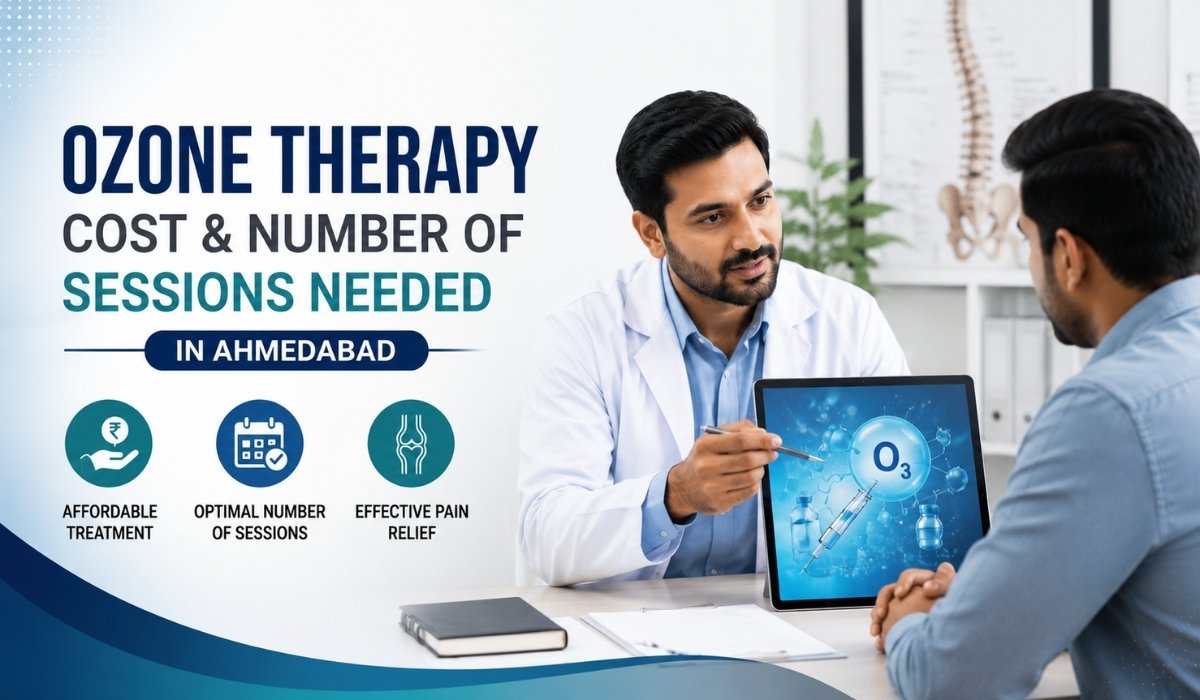
Ozone Therapy Cost & Number of Sessions Needed in Ahmedabad

Doctor explaining ozone therapy treatment cost and number of sessions for pain management in Ahmedabad

Quick Answer

• In Ahmedabad, a single ozone therapy session for pain management typically costs between ₹2,000 and ₹6,000, depending on the injection site, the volume of ozone-oxygen mixture used, and whether imaging guidance (X-ray or ultrasound) is involved.

• Most patients need 3 to 6 sessions spaced 1–2 weeks apart for joint or muscle pain, and 4 to 8 sessions for disc-related back and sciatica pain, though your treating specialist decides the exact number based on your MRI findings and response to early sessions.

• Ozone therapy is generally more affordable than surgery and often comparable to or slightly higher than a single epidural injection, but total cost depends on how many sessions your specific condition requires.

Quick Summary 

  • Per-session cost in Ahmedabad usually falls between ₹2,000–₹6,000 depending on the site treated and imaging guidance used.
  • Total treatment cost ranges roughly ₹8,000–₹35,000 across a full protocol, not a single visit.
  • Most musculoskeletal and joint conditions need 3–6 sessions; disc-related back pain often needs 4–8.
  • Sessions are typically spaced 1–2 weeks apart to let the anti-inflammatory effect build.
  • Ozone therapy is not a permanent cure — it is one tool within a broader, evidence-based pain management plan.
  • Response is evaluated after the first 2–3 sessions; the specialist may adjust the plan rather than committing to a fixed package upfront.

Introduction

If you’re researching ozone therapy for back pain, sciatica, or joint pain in Ahmedabad, two questions usually come first: what will it cost, and how many sessions will you actually need? The honest answer is that both depend heavily on what’s causing your pain, how long you’ve had it, and how your body responds to the first few sessions — which is why credible clinics avoid quoting a flat number before an assessment.

This guide breaks down real-world pricing patterns for ozone therapy in Ahmedabad, the factors that move the cost up or down, typical session counts for common conditions, and how ozone compares to other non-surgical options like PRP and radiofrequency ablation. The goal is to help you budget realistically and ask your specialist the right questions before you begin.

What Is Ozone Therapy and How Is It Used for Pain?

Ozone therapy for pain management involves injecting a precise, medical-grade mixture of ozone and oxygen gas into or around a painful joint, muscle, or the space near a spinal disc. The goal is to reduce local inflammation, improve oxygen delivery to the tissue, and calm the irritated nerve signals that produce pain.

It is used as a non-surgical option for conditions such as chronic low back pain, sciatica from a mildly herniated disc, knee and shoulder joint pain, and some cases of tendon-related pain, usually after simpler measures like physiotherapy and medication have been tried first.

Expert Insight

• Ozone is not injected randomly — it is delivered to a specific ‘pain generator’ (the disc, joint, or muscle believed to be the source), often under X-ray or ultrasound guidance for accuracy. This precision is one reason cost varies: a guided intradiscal injection costs more than a simple intramuscular one.

What Does Ozone Therapy Cost in Ahmedabad?

Across India, published pricing for ozone therapy sessions ranges widely — anywhere from a few hundred rupees for a simple intramuscular pass at a general wellness centre, up to ₹8,000–₹10,000 for advanced protocols like extracorporeal blood ozonation (EBOO) at specialty hospitals. For targeted pain-management use at an interventional pain clinic in Ahmedabad, most patients should budget in the ₹2,000–₹6,000 per session range.

What Changes the Price Per Session

  • Injection site: a single joint injection typically costs less than an image-guided spinal/paraspinal injection.
  • Imaging guidance: sessions performed under C-arm X-ray or ultrasound guidance cost more than blind injections, but are safer and more accurate.
  • Ozone concentration and volume: higher concentrations or larger treated volumes increase consumable cost.
  • Number of sites treated in one visit: treating both the disc and surrounding paraspinal muscles in one sitting costs more than a single site.
  • Clinic setting: hospital-based interventional suites generally cost more than smaller outpatient setups, reflecting sterility standards and monitoring.
Treatment TypeTypical Cost Range (per session)Typical Sessions Needed
Intramuscular/paraspinal ozone injection₹2,000 – ₹3,5003 – 6 sessions
Ozone injection into a peripheral joint (knee, shoulder)₹2,500 – ₹4,5003 – 5 sessions
Image-guided perinuclear/intradiscal ozone injection₹4,000 – ₹6,5001 – 3 sessions
Ozone combined with PRP in the same sitting₹6,000 – ₹10,0003 – 4 sessions

A full course of ozone therapy for a disc-related back pain condition — including consultation, imaging review, and 4–6 sessions — commonly totals somewhere between ₹15,000 and ₹35,000 in Ahmedabad. This is usually less than the cost of spine surgery and its associated hospital stay, though it is not a like-for-like substitute in every case; your specialist will tell you if your condition is actually a surgical one.

How Many Ozone Therapy Sessions Will You Actually Need?

There is no universal number, and any clinic that promises an exact session count before examining you or reviewing your MRI is skipping an important step. That said, published protocols and typical clinical practice give a reasonable range to plan around.

ConditionTypical Session RangeTypical Interval Between Sessions
Chronic low back pain (muscular/facet-related)3 – 5 sessions1 week
Sciatica from mild-to-moderate disc herniation4 – 8 sessions1 – 2 weeks
Knee osteoarthritis / joint pain3 – 6 sessions1 week
Frozen shoulder / tendon-related pain3 – 5 sessions1 – 2 weeks
Post-surgical or complex chronic pain6 – 10 sessions (reviewed periodically)1 – 2 weeks


Real-Life Scenario

• A 45-year-old office worker with sciatica from a mild L5-S1 disc bulge typically starts with 2 image-guided ozone sessions two weeks apart. If pain drops by 50% or more, the specialist may add 2 more sessions to consolidate the improvement; if response is minimal, imaging is reviewed again before continuing rather than repeating the same protocol indefinitely.

Clinics that follow evidence-based protocols reassess after the first 2–3 sessions rather than locking patients into a fixed 10-session package upfront. This protects you from paying for sessions that aren’t producing measurable improvement.

Ozone Therapy vs Other Non-Surgical Pain Options: Cost Perspective

TreatmentTypical Cost (Ahmedabad)Best Suited For
Ozone therapy₹2,000 – ₹6,500 per sessionMild-moderate disc pain, joint pain, muscular pain
Epidural steroid injection₹8,000 – ₹18,000 per sessionRadicular pain with significant nerve compression
PRP therapy₹6,000 – ₹15,000 per sessionTendon, ligament, and early joint degeneration
Radiofrequency ablation (RFA)₹25,000 – ₹60,000 per procedureFacet-joint driven chronic back/neck pain

Ozone therapy generally sits at the more affordable end of interventional pain options, which is one reason it’s often tried before more invasive or expensive procedures — provided imaging and clinical examination confirm it’s an appropriate fit for the specific pain source.

Is Ozone Therapy Safe? What to Know Before You Budget for It

When performed by a trained interventional pain specialist under proper imaging guidance and sterile technique, ozone therapy is generally well tolerated. Mild soreness at the injection site for a day or two is common. Serious complications are rare but not zero, which is why guided, protocol-based administration by a qualified specialist matters more than the lowest quoted price.

Common Mistakes to Avoid

• Choosing a provider based purely on the lowest per-session price without checking whether imaging guidance is included.

• Expecting a single session to resolve chronic, long-standing pain — most protocols need a course, not one visit.

• Stopping physiotherapy or activity modification because ozone injections are ‘handling it’ — ozone works best alongside rehabilitation, not instead of it.

• Not asking what happens if the first 2–3 sessions show minimal improvement — a good clinic has a clear next step, not just ‘more sessions.’

Myths vs Facts: Ozone Therapy Cost & Treatment

MythFact
Cheaper ozone therapy always means the same treatment at a lower price.Price differences often reflect real differences in imaging guidance, ozone concentration, and physician experience, not just markup.
More sessions always mean better results.Response should be reassessed every 2–3 sessions; continuing without improvement wastes money and delays other options.
Ozone therapy replaces the need for physiotherapy.It works best as part of a combined plan that includes rehabilitation and activity modification.
Ozone therapy is a permanent cure for disc herniation.It reduces inflammation and pain in a large share of appropriately selected patients, but it manages the condition rather than curing the underlying disc pathology.

Questions Patients Forget to Ask Before Starting

  1. Is imaging guidance (X-ray or ultrasound) included in the quoted price, or billed separately?
  2. How will we know after 2–3 sessions whether the treatment is working?
  3. What is the plan if my pain doesn’t improve after the initial sessions?
  4. Are there any activities I should avoid or modify after the treatment?
  5. Who is administering the injection — an interventional pain specialist or general practitioner?

Practical Action Plan

  1. Get a clear diagnosis first — an MRI or clinical exam should confirm ozone is appropriate for your specific pain source.
  2. Ask for a written estimate covering the likely number of sessions, not just the per-session price.
  3. Confirm whether imaging guidance is used and included in the cost.
  4. Track your pain and function after each session so you and your specialist can objectively judge progress.
  5. Pair sessions with physiotherapy or activity modification for a more durable result.

Nexus Pain Expert Summary

Nexus Advanced Pain Management — Expert Guidance

• At Nexus Advanced Pain Management in Ahmedabad, ozone therapy is offered as part of a comprehensive pain assessment — not as a standalone package sold before diagnosis.

• Treatment plans are built around imaging findings and reviewed every few sessions so patients aren’t paying for injections that aren’t producing results.

• Ozone therapy is used alongside rehabilitation guidance and, where appropriate, combined with other evidence-based non-surgical options for a more complete recovery plan.

Key Takeaways

  • Expect ₹2,000–₹6,500 per session in Ahmedabad, with total course costs typically ₹15,000–₹35,000.
  • Most patients need 3–8 sessions depending on the condition, spaced 1–2 weeks apart.
  • Image-guided injections cost more but are generally more precise, especially for disc-related pain.
  • Response should be reviewed every 2–3 sessions rather than committing to a long fixed package upfront.
  • Ozone therapy works best combined with physiotherapy, not as a replacement for it.
  • Always confirm your diagnosis and treatment rationale with a qualified interventional pain specialist before starting.

How Nexus Pain Can Help

Nexus Advanced Pain Management approaches ozone therapy as one option within a comprehensive, evidence-based pain assessment. This includes a detailed clinical evaluation, review of relevant imaging, personalized treatment planning, and — where ozone therapy is appropriate — image-guided administration by an interventional pain specialist.

Treatment plans are paired with rehabilitation guidance and long-term pain management strategies rather than a one-off injection, with progress reviewed at regular intervals so the plan can be adjusted based on how each patient actually responds.

Frequently Asked Questions

Is ozone therapy covered by health insurance in India?

Coverage varies by insurer and policy; many treat it as a non-surgical/OPD procedure that may not be fully covered. Check directly with your insurer before starting treatment.

How long does one ozone therapy session take?

Most sessions take 15 to 45 minutes, including preparation and post-procedure observation, and are done on an outpatient basis without anesthesia.

Can ozone therapy be combined with physiotherapy?

Yes — combining ozone injections with structured physiotherapy is common practice and often supports better, more lasting outcomes than injections alone.

How soon will I feel pain relief after an ozone injection?

Some patients notice improvement within a few days, while for others it builds gradually over 2–3 sessions as inflammation reduces.

Does a higher price always mean a better ozone therapy provider?

Not necessarily, but very low prices can signal that imaging guidance, sterile protocol standards, or specialist experience are being cut. Ask what’s included before comparing quotes.

Is ozone therapy painful?

Most patients report mild discomfort similar to a routine injection; some soreness at the site for a day or two afterward is common.

Can ozone therapy be used for sciatica?

Yes, ozone injections near the affected disc or nerve root are commonly used for sciatica caused by mild-to-moderate disc herniation.

What happens if ozone therapy doesn’t work for me?

A good specialist reassesses after a few sessions and discusses alternative options such as PRP, epidural injections, or radiofrequency ablation rather than repeating an ineffective plan.

Are there age restrictions for ozone therapy?

There’s no strict age cutoff, but suitability depends on overall health, the specific condition being treated, and any contraindications identified during assessment.

How many total sessions are typical for chronic back pain?

Most protocols for chronic low back pain range from 4 to 8 sessions, with progress reviewed periodically rather than fixed in advance.

Can I return to work the same day after a session?

Most patients resume light activity the same day or the next, though your specialist may recommend a brief rest period depending on the injection site.

Where can I get ozone therapy for pain management in Ahmedabad?

Interventional pain clinics such as Nexus Advanced Pain Management in Ahmedabad offer ozone therapy as part of an assessed, non-surgical pain management plan.


Categories Shoulder Pain, Frozen Shoulder

Frozen Shoulder vs Shoulder Injury: Understanding the Difference

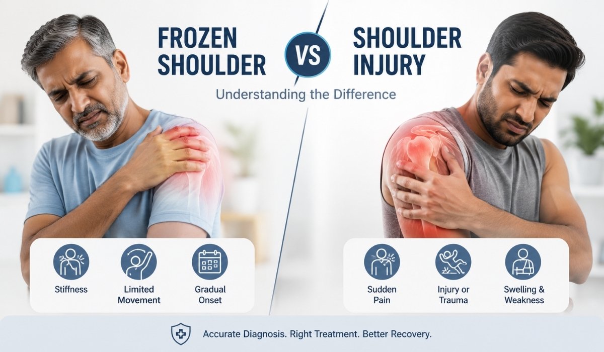
Illustration comparing frozen shoulder and shoulder injury, highlighting symptoms, pain areas, and limited shoulder movement.

Quick Answer

Frozen shoulder (adhesive capsulitis) is a stiffening and thickening of the shoulder joint capsule that restricts movement in every direction, including when someone else moves your arm for you. A shoulder injury — such as a rotator cuff tear, impingement, or strain — is damage to a specific muscle, tendon, or structure that usually causes pain and weakness with active movement, while passive movement (someone else lifting your arm) is often still possible. The passive range-of-motion test is the single biggest clue that separates the two.

Quick Summary

  • Frozen shoulder restricts movement in all directions, even with assistance; most shoulder injuries allow more passive movement than active movement.
  • Frozen shoulder usually develops gradually over weeks to months; injuries like rotator cuff tears can happen suddenly from a fall or lifting strain, or build up over years of overuse.
  • Frozen shoulder affects roughly 2–5% of the general population and is more common between ages 40–60, particularly in women and people with diabetes.
  • A rotator cuff tear can cause sharp pain and noticeable weakness, especially when raising the arm overhead; frozen shoulder pain is typically deep, diffuse, and worse at night.
  • Frozen shoulder progresses through three stages — freezing, frozen, and thawing — that can span months to a couple of years.
  • Imaging (ultrasound or MRI) helps confirm a structural injury and can also identify capsular thickening consistent with frozen shoulder.
  • Frozen shoulder is overwhelmingly managed without surgery, while some rotator cuff tears may eventually need surgical repair if conservative care fails.

Introduction

Shoulder pain is one of the most common reasons people in Ahmedabad visit a pain management clinic, and one of the most common points of confusion is whether the problem is a frozen shoulder or an underlying shoulder injury. Both can cause pain, stiffness, and difficulty sleeping on the affected side, but the underlying mechanism, expected recovery path, and treatment approach are quite different. Getting the distinction right early matters — treating a rotator cuff tear like a frozen shoulder, or vice versa, can delay real recovery by months.

This guide breaks down how frozen shoulder and shoulder injuries differ in symptoms, causes, diagnosis, and treatment, so you know what questions to ask before your consultation.

What Is Frozen Shoulder?

Frozen shoulder, medically known as adhesive capsulitis, occurs when the capsule of connective tissue surrounding the shoulder joint becomes inflamed, thickens, and tightens. This shrinks the space available for the joint to move, restricting motion in every direction — forward, backward, and sideways — regardless of whether the movement is active (done by the patient) or passive (assisted by someone else).

Expert Insight: In clinical practice, frozen shoulder is often missed in its early “freezing” stage because pain, not stiffness, is the dominant complaint at first. Patients frequently assume it’s a muscle strain and delay assessment until motion loss becomes obvious — by which point the stiffer “frozen” stage has already set in.

What Is a Shoulder Injury?

“Shoulder injury” is a broad term covering damage to a specific structure in the shoulder — most often the rotator cuff tendons, but also the labrum, bursa, or surrounding muscles. Common types include rotator cuff tears (partial or complete), shoulder impingement, tendinitis, and bursitis. Injuries can be acute (a fall, a heavy lift, a sports collision) or degenerative (built up gradually from repetitive overhead work, age-related wear, or occupations like painting, carpentry, or racquet sports).

Unlike frozen shoulder, most shoulder injuries leave passive range of motion relatively intact — a clinician or family member can often lift the arm through most of its normal arc, even though the patient cannot do it unassisted due to pain or weakness.

Symptoms: How to Tell Them Apart

The clearest bedside clue is the passive range-of-motion test: if someone else can lift your arm for you through a near-normal arc, an injury such as a rotator cuff tear is more likely. If the shoulder feels physically “locked” even with assistance, frozen shoulder is more likely.

FeatureFrozen ShoulderShoulder Injury (e.g. Rotator Cuff Tear)
OnsetGradual, over weeks to monthsOften sudden (fall, lift) or gradual overuse
Passive motionRestricted even with assistanceOften preserved with assistance
Pain characterDeep, diffuse ache, worse at nightSharp pain, often with specific movements
WeaknessUncommon as a primary symptomCommon, especially lifting overhead
ProgressionThree stages over months to ~2 yearsVaries; can worsen without treatment
Typical age group40–60 years, more common in womenAny age; higher in athletes and manual workers

Causes

Frozen shoulder causes include:

  • Idiopathic onset (no clear trigger) — the most common pattern
  • Prolonged immobilization after a prior injury, fracture, or surgery
  • Diabetes and thyroid disorders, which significantly raise risk
  • Underlying shoulder inflammation left untreated

Shoulder injury causes include:

  • Acute trauma — a fall onto an outstretched arm, sudden heavy lifting, or a sports collision
  • Repetitive overhead motion — swimming, painting, racquet sports, manual labour
  • Age-related degeneration of tendon tissue, which raises tear risk after the mid-40s
  • Poor posture and muscle imbalance contributing to impingement

Risk Factors

  • Age 40–60 (frozen shoulder); any age with repetitive strain (injuries)
  • Diabetes, thyroid disease, or cardiac conditions (frozen shoulder)
  • Recent shoulder or arm surgery, stroke, or a period of arm immobilisation
  • Occupations or sports requiring repetitive overhead arm movement
  • Previous shoulder injury that was not fully rehabilitated

Diagnosis

A thorough clinical examination is usually the starting point — comparing active versus passive range of motion, checking for specific weakness patterns, and reviewing how the pain began. Imaging plays a confirming role: an MRI or ultrasound can rule out or confirm a structural tear, and can occasionally show the capsular thickening characteristic of frozen shoulder. X-rays are sometimes used to rule out arthritis or bone spurs contributing to impingement.

Common Mistake: Self-diagnosing based on pain location alone. Pain in the same general area of the shoulder can come from the capsule, the tendons, the bursa, or even referred pain from the neck — a proper assessment is what actually separates these conditions.

Treatment Options

Treatment approaches diverge meaningfully once a diagnosis is confirmed.

Frozen ShoulderShoulder Injury
Physiotherapy focused on gentle, progressive stretchingPhysiotherapy focused on strengthening and controlled loading
Anti-inflammatory pain managementAnti-inflammatory pain management
Corticosteroid or hydrodilatation injections in select casesTargeted injections or PRP therapy for tendon healing
Radiofrequency ablation for select chronic pain casesRadiofrequency ablation for select chronic pain cases
Surgery rarely neededSurgery considered if conservative care fails or tear is complete

Non-Surgical Treatment Approaches

Most frozen shoulder cases, and a large share of shoulder injuries, respond well to non-surgical, interventional pain management. Structured physiotherapy, targeted injections, and image-guided procedures such as radiofrequency ablation or PRP therapy can meaningfully reduce pain and restore function without the recovery time associated with surgery. The right combination depends on the specific diagnosis, severity, and how long symptoms have been present.

Expert Insight: The earlier a frozen shoulder is correctly identified, the more responsive it tends to be to conservative treatment. Waiting through the freezing stage without intervention often means a longer, stiffer frozen stage before recovery begins.

Prevention Tips

  • Keep the shoulder moving through its normal range even after a minor injury — avoid unnecessary prolonged immobilisation
  • Manage diabetes and thyroid conditions proactively, as these directly raise frozen shoulder risk
  • Warm up before overhead activity and build shoulder and rotator cuff strength gradually
  • Seek assessment for shoulder pain that persists beyond 2–3 weeks rather than waiting it out
  • Complete any prescribed physiotherapy fully, even once pain improves, to avoid re-injury or stiffness

Recovery and Rehabilitation

Frozen shoulder recovery is typically the slower of the two, often spanning several months to around two years across its freezing, frozen, and thawing stages, though most patients see steady improvement with consistent physiotherapy. Shoulder injury recovery timelines vary more widely — mild strains and tendinitis often improve within 4–8 weeks of guided rehabilitation, while a significant rotator cuff tear, if treated conservatively, may take 3–6 months to regain full strength, and longer if surgery is required.

Common Mistake: Stopping rehabilitation exercises as soon as pain eases. Pain relief is not the same as full recovery — stopping early is one of the most frequent reasons stiffness or weakness returns.

Expert Insight: When Both Conditions Overlap

It is possible to have a rotator cuff injury and later develop frozen shoulder, particularly if the arm was kept still for an extended period during recovery. This is why ongoing follow-up matters even after an initial injury has been treated.

Common Mistakes Patients Make

  • Assuming all shoulder pain is “just a strain” and delaying evaluation for months
  • Relying only on painkillers without addressing the underlying stiffness or structural cause
  • Over-resting the shoulder for frozen shoulder, which can worsen stiffness rather than help it

Myths vs Facts

MythFact
Frozen shoulder always needs surgery.Most cases improve with physiotherapy and non-surgical pain management; surgery is uncommon.
If you can move your arm at all, it can’t be frozen shoulder.Some limited active movement is still possible, especially in early stages; the key marker is restricted passive movement.
Rest is always best for shoulder pain.Complete immobilisation can worsen or trigger frozen shoulder; guided, gentle movement is usually recommended instead.
A rotator cuff tear always requires surgery.Many tears, especially partial ones, respond well to physiotherapy and interventional treatment first.

Questions Patients Forget to Ask

  • Is my shoulder pain coming from the joint capsule, a tendon, or somewhere else, like my neck?
  • What stage is my frozen shoulder in, and how does that change my treatment plan?
  • Will imaging actually change my treatment, or is a clinical exam enough for now?
  • What specific home exercises are safe for my exact diagnosis, and which should I avoid?
  • At what point would we consider an injection, PRP, or radiofrequency ablation instead of continuing physiotherapy alone?

Practical Action Plan

  1. If shoulder pain or stiffness has lasted more than 2–3 weeks, book a clinical assessment rather than waiting it out.
  2. Note whether someone else can lift your arm for you comfortably — this detail helps guide the initial diagnosis.
  3. Avoid prolonged immobilisation; keep the shoulder gently mobile unless specifically advised otherwise.
  4. Ask your clinician whether imaging is needed to confirm a structural injury or capsular thickening.
  5. Follow the full physiotherapy course prescribed, even after pain starts improving.
  6. Discuss non-surgical options — physiotherapy, injections, PRP, or radiofrequency ablation — before considering surgery.

Nexus Pain Expert Summary Box

Distinguishing frozen shoulder from a shoulder injury starts with a comprehensive pain assessment that looks at active versus passive movement, pain pattern, and relevant risk factors such as diabetes or a prior injury. Based on that assessment, an evidence-based, personalised treatment plan can combine physiotherapy guidance, non-surgical interventional pain procedures, and long-term rehabilitation support — with the goal of restoring shoulder function without unnecessary surgery wherever possible. Lifestyle and movement guidance is also provided to reduce the chance of recurrence.

Key Takeaways

  • Frozen shoulder restricts both active and passive movement; most injuries mainly limit active movement.
  • Onset is gradual for frozen shoulder, often sudden for acute injuries like tears.
  • Diabetes, thyroid conditions, and prior immobilisation raise frozen shoulder risk.
  • A proper clinical exam, and imaging when needed, is the reliable way to tell the two apart.
  • Both conditions usually respond to non-surgical, physiotherapy-led treatment when caught early.
  • Delaying assessment tends to prolong recovery for either condition.
  • Recovery timelines differ significantly, so an accurate diagnosis sets realistic expectations.

Frequently Asked Questions

  1. What is the main difference between frozen shoulder and a shoulder injury?

Frozen shoulder restricts movement in every direction, even when someone else moves your arm for you, because the joint capsule itself has tightened. A shoulder injury, such as a rotator cuff tear, usually still allows passive movement even when active movement is painful or weak.

  1. Can a shoulder injury turn into frozen shoulder?

Yes. Prolonged immobilisation after an injury or surgery is a recognised risk factor for developing frozen shoulder afterward, which is why gentle, guided movement during recovery matters.

  1. How long does frozen shoulder usually last?

Frozen shoulder typically progresses through freezing, frozen, and thawing stages over several months to around two years, though consistent physiotherapy can support steadier improvement.

  1. Is frozen shoulder more common in people with diabetes?

Yes, diabetes is a well-established risk factor for frozen shoulder, along with thyroid disorders and periods of shoulder immobilisation.

  1. Does a rotator cuff tear always need surgery?

Not always. Many tears, particularly partial ones, are managed successfully with physiotherapy and non-surgical pain management before surgery is considered.

  1. What does the “passive range of motion” test actually check?

It checks whether someone else can move your arm through its normal range for you. Preserved passive motion points more toward an injury like a tear; restricted passive motion points more toward frozen shoulder.

  1. Can I have both a rotator cuff injury and frozen shoulder at the same time?

Yes, this combination does occur, which is one reason accurate clinical assessment is important rather than assuming just one condition is present.

  1. Is shoulder pain that’s worse at night more typical of frozen shoulder or an injury?

Night pain is common in both, but the deep, diffuse ache of frozen shoulder disrupting sleep is a classic pattern, while injury-related night pain often relates to specific sleeping positions that load the tendon.

  1. What tests are used to diagnose frozen shoulder versus a shoulder injury?

A clinical exam comparing active and passive movement is the starting point; ultrasound or MRI can then confirm a structural tear or show capsular thickening if the diagnosis remains unclear.

  1. Is exercise safe for frozen shoulder, or should the shoulder be rested?

Gentle, guided movement is generally recommended over complete rest, since prolonged immobilisation can worsen stiffness. Exercises should be tailored to your specific stage and diagnosis.

  1. When should I see a pain management specialist for shoulder pain?

If shoulder pain or stiffness has persisted beyond 2–3 weeks, is limiting daily activities, or is affecting sleep, it’s worth getting a clinical assessment rather than waiting for it to resolve on its own.

  1. Where can I get frozen shoulder or shoulder injury treatment in Ahmedabad?

Nexus Pain Management in Ahmedabad offers comprehensive shoulder pain assessment and evidence-based, non-surgical treatment planning, including physiotherapy guidance and interventional pain procedures where appropriate.

Categories PRP Therapy

Is PRP Effective for Knee Arthritis?

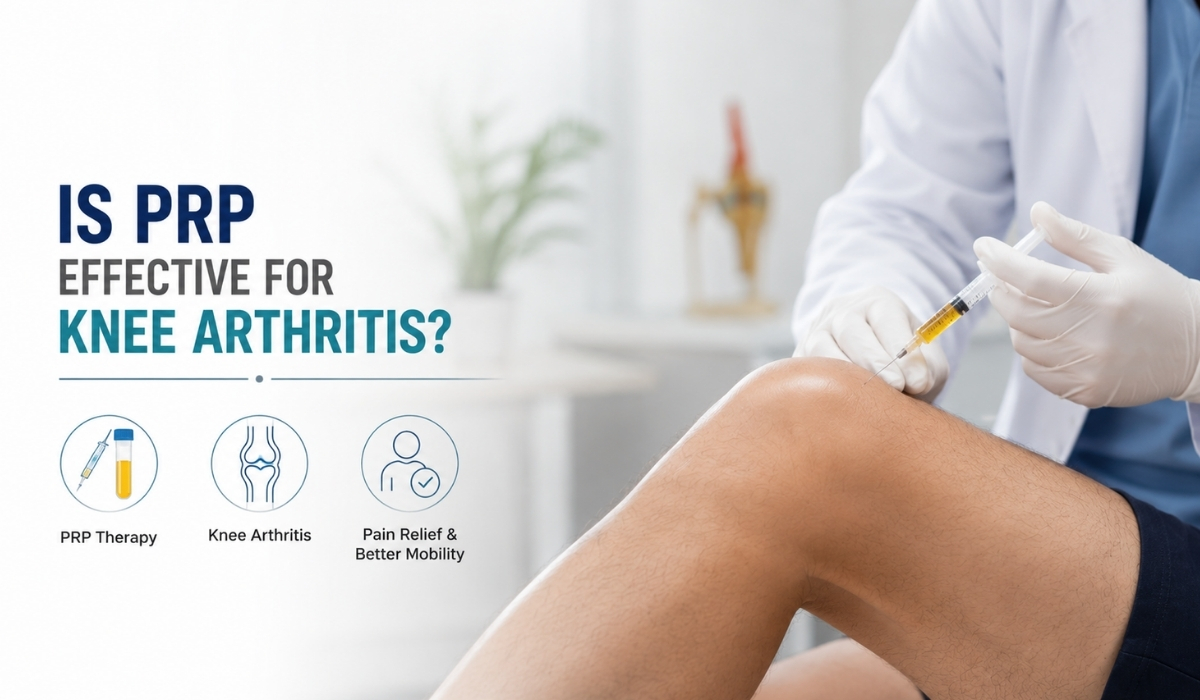
Prp theraphy for Knee Arthritis

Quick Answer

•     Yes — for mild to moderate knee osteoarthritis (Kellgren-Lawrence grade I–III), current research shows PRP gives better and longer-lasting pain and function improvement than hyaluronic acid or corticosteroid injections.

•     PRP works best as an early-stage, symptom-management option — not a cure — and typically needs a series of 2–3 injections for the strongest results.

•     It is less effective in advanced, bone-on-bone arthritis, where structural support options are usually more appropriate.

Quick Summary

  • PRP uses concentrated platelets from your own blood to calm knee inflammation and support the joint’s healing response.
  • 2024–2025 meta-analyses report PRP outperforming hyaluronic acid and corticosteroids for pain relief and function in knee osteoarthritis.
  • Best results are seen in mild-to-moderate osteoarthritis; benefit drops in severe, bone-on-bone joints.
  • A single injection helps some patients, but a series of 2–3 injections spaced a few weeks apart tends to work meaningfully better.
  • Relief commonly lasts 6–12 months, and some patients report benefit up to 24 months.
  • Major bodies such as the American College of Rheumatology and OARSI still stop short of formally endorsing PRP, mainly because preparation protocols vary so much between clinics.
  • Higher BMI and a longer history of arthritis are linked to weaker results — earlier treatment tends to help more.
  • PRP does not reverse advanced joint damage; it manages symptoms and may help delay other interventions.

Introduction

If you’ve been told PRP therapy could help your knee arthritis, you’ve probably also come across conflicting opinions — some call it a breakthrough, others call it overhyped. The honest answer sits in between. Platelet-rich plasma (PRP) is not a miracle cure, but a growing body of clinical research, including several 2024–2025 meta-analyses, shows it can meaningfully reduce knee arthritis pain and improve function for the right patient. This guide walks through what PRP actually is, what the evidence says about its effectiveness, who benefits most, and what to realistically expect from treatment.

What Is PRP Therapy?

Platelet-rich plasma is prepared by drawing a small sample of your own blood, spinning it in a centrifuge to concentrate the platelets, and injecting that concentrate directly into the knee joint under sterile, often ultrasound-guided, conditions. Platelets contain growth factors that play a role in tissue repair and modulating inflammation, which is the biological rationale for using PRP in osteoarthritic joints.

Because PRP is made from the patient’s own blood, it carries a low risk of allergic reaction, which is one reason it has become a widely used non-surgical option for knee pain over the last decade.

How PRP Works for an Arthritic Knee

Knee osteoarthritis involves progressive cartilage wear, low-grade joint inflammation, and changes in the joint fluid that normally cushions and lubricates movement. PRP is thought to work through two main mechanisms:

  • Anti-inflammatory effect: growth factors and signalling proteins in platelets can help reduce inflammatory activity inside the joint, which is a major driver of arthritis pain.
  • Supportive tissue response: PRP may help support the joint’s natural repair processes and improve the quality of joint fluid, though it does not regenerate lost cartilage in advanced disease.

Is PRP Effective for Knee Arthritis? What the Evidence Actually Shows

This is the question most patients really want answered, so here is a straightforward summary of where the research currently stands.

What multiple 2024–2025 studies agree on

  • PRP — particularly leukocyte-poor PRP — shows superior pain relief and functional improvement compared with hyaluronic acid and corticosteroid injections, especially in mild-to-moderate osteoarthritis (Kellgren-Lawrence grades I–III).
  • A large meta-analysis found the most significant improvement at the 6-month mark, with benefits considered clinically meaningful, not just statistically significant.
  • A 2025 review comparing PRP to hyaluronic acid, corticosteroids, and bone marrow aspirate concentrate found PRP performs better than HA and steroids, and comparably to bone marrow concentrate.
  • A course of injections works better than a single shot — one retrospective study found a series was roughly four times more effective for pain relief than one injection alone.

Where caution is still warranted

  • Major professional bodies, including the American College of Rheumatology and OARSI, have not formally endorsed PRP for knee osteoarthritis, largely because PRP preparation protocols vary widely between clinics and studies, making outcomes harder to standardise and compare.
  • Patients with a higher BMI or a longer-standing history of arthritis tend to see smaller benefits — earlier-stage treatment generally performs better.
  • PRP is less effective in advanced, bone-on-bone osteoarthritis, where little cushioning cartilage remains.
Expert Insight:

At Nexus Pain, we treat PRP as a genuinely useful tool in the mild-to-moderate osteoarthritis stage — not as a universal fix. The patients who respond best are typically those who start treatment before the joint reaches advanced, bone-on-bone changes.

PRP vs Hyaluronic Acid vs Corticosteroids

FactorPRPHyaluronic AcidCorticosteroids
Pain relief duration6–12 months, sometimes up to 24Typically 3–6 monthsWeeks to a few months
Best-suited stageMild to moderate OAMild to moderate OAShort-term flare-ups
MechanismAnti-inflammatory + tissue supportJoint lubricationStrong anti-inflammatory
Repeat use concernGenerally well toleratedWell toleratedRepeated use may affect cartilage over time
Evidence strength (2024–25)Moderate, growing, favourableEstablished but modest effectEstablished, short-lived effect

Who Is a Good Candidate for PRP?

PRP tends to work best for a fairly specific patient profile, and being upfront about this is part of setting realistic expectations.

  • Diagnosed with mild-to-moderate knee osteoarthritis (Kellgren-Lawrence grade I–III)
  • Wants to reduce reliance on repeated corticosteroid shots or delay surgery
  • Has a reasonably active lifestyle and wants to preserve joint function
  • Does not have an active infection, blood disorder, or certain cancers (screened during assessment)

Patients with advanced, bone-on-bone arthritis are usually better served by discussing structural or surgical options, since PRP has limited cartilage left to support at that stage.

The PRP Procedure: What to Expect

  1. A small blood sample is drawn, similar to a routine blood test.
  2. The sample is processed in a centrifuge to separate and concentrate the platelets.
  3. Under sterile technique — often with ultrasound guidance for accuracy — the concentrated PRP is injected into the knee joint.
  4. The full procedure typically takes 30–45 minutes, and most patients go home the same day.
  5. A course of 2–3 injections, spaced roughly 2–4 weeks apart, is a common protocol used to improve outcomes.

Recovery and What Happens After

Mild soreness, swelling, or stiffness at the injection site for one to three days is the most common after-effect — this is a normal part of the inflammatory response PRP relies on, not a complication. Most patients resume light daily activity within a day or two, while higher-impact activity is usually paced back in over one to two weeks, guided by your treating physician.

  • Days 1–3: mild soreness/swelling is normal; ice and rest as advised
  • Week 1–2: gradual return to daily activities
  • Weeks 3–6: functional improvement often becomes noticeable
  • Months 3–6: peak benefit typically reported in current research

Supporting Your Results: Lifestyle Factors That Matter

Because outcomes are linked to body weight and disease duration, a few supporting habits genuinely influence how well PRP performs.

  • Maintaining a healthy body weight to reduce load on the knee joint
  • Low-impact strengthening (physiotherapy-guided) to support joint stability
  • Addressing knee pain early rather than waiting until arthritis has progressed
  • Avoiding repeated high-impact stress on the joint during the healing window after injection

Expert Insight Sections

Expert Insight 1:

Patients often ask if PRP can “regrow” cartilage. It cannot reverse structural cartilage loss in advanced arthritis — its real value is calming inflammation and supporting joint function in the earlier stages of disease.

Expert Insight 2:

The gap between a single injection and a full course (2–3 injections) is one of the most consistent findings in recent research — patients considering PRP should ask their clinic about a series-based protocol rather than a one-time shot.

Expert Insight 3:

Not all PRP is prepared the same way. Platelet concentration and leukocyte content vary by clinic, and this variation is part of why professional bodies remain cautious — ask your provider how your PRP is prepared and processed.

Common Mistakes Patients Make

  • Expecting PRP to reverse advanced, bone-on-bone arthritis rather than manage early-to-moderate disease
  • Choosing a single injection to save cost, then judging PRP as “ineffective” when a course was actually indicated
  • Returning to high-impact activity too soon after injection instead of following a gradual, guided return

Myths vs Facts

MythFact
PRP cures knee arthritis permanently.PRP manages symptoms and inflammation; it does not reverse underlying joint damage.
One PRP shot is enough for lasting results.Research suggests a series of 2–3 injections generally outperforms a single injection.
PRP works the same at every clinic.Preparation protocols (platelet and leukocyte content) vary and directly affect outcomes.
PRP is only for athletes.It is used across age groups, most commonly for adults with mild-to-moderate osteoarthritis.

Questions Patients Forget to Ask

  • What grade is my knee osteoarthritis, and does that make me a good PRP candidate?
  • How is your clinic’s PRP prepared — platelet-rich, leukocyte-poor, or leukocyte-rich?
  • Will I need a single injection or a course, and how is that decided?
  • What realistic improvement, in pain and function, can I expect by month three and month six?
  • What happens if PRP doesn’t give me enough relief — what’s the next step?

Practical Action Plan

  1. Get an accurate diagnosis and osteoarthritis grading (clinical exam plus imaging) before considering PRP.
  2. Discuss whether your stage of arthritis fits the profile that responds best to PRP.
  3. Ask about the clinic’s PRP preparation protocol and whether a single injection or a course is recommended for your case.
  4. Set a realistic 3–6 month timeline to judge results, rather than expecting immediate change.
  5. Pair treatment with weight management and guided low-impact strengthening for better odds of a good outcome.

Nexus Pain Expert Summary Box

Nexus Pain’s Perspective:

PRP is one option within a broader non-surgical pain management plan, not a stand-alone fix. At Nexus Pain Management, PRP is considered after a full assessment of your osteoarthritis stage, alongside physiotherapy, activity modification, and — where appropriate — other interventional options, so the treatment plan matches the actual condition of your knee rather than a one-size-fits-all protocol.

Key Takeaways

  • PRP has moderate, growing evidence supporting its effectiveness for mild-to-moderate knee osteoarthritis.
  • It generally outperforms hyaluronic acid and corticosteroids for pain relief duration and functional improvement.
  • A course of injections tends to outperform a single injection.
  • Results are weaker in advanced, bone-on-bone arthritis and in patients with higher BMI or long-standing disease.
  • Major medical bodies remain cautious due to inconsistent preparation protocols across providers.
  • PRP works best as part of a broader, personalised pain management plan — not a stand-alone cure.

How Nexus Pain Can Help

Nexus Pain Management approaches knee arthritis with a comprehensive pain assessment before recommending any treatment, including PRP. This includes evidence-based, personalised treatment planning, non-surgical pain management options, and — where clinically appropriate — interventional pain procedures. The focus throughout is on realistic, long-term pain relief strategies, rehabilitation guidance, and lifestyle modification support, so each treatment plan reflects the actual stage and needs of the patient’s knee, not a generic protocol.

Frequently Asked Questions

1. Is PRP actually effective for knee arthritis?

For mild-to-moderate knee osteoarthritis, current research shows PRP can offer meaningful pain relief and functional improvement, generally outperforming hyaluronic acid and corticosteroid injections in recent studies.

2. How many PRP injections do I need for knee arthritis?

A course of 2–3 injections spaced a few weeks apart is a common protocol and tends to produce better results than a single injection.

3. How long does PRP relief last?

Many patients report benefit for 6–12 months, with some studies noting improvement lasting up to 24 months, particularly in earlier-stage arthritis.

4. Does PRP regrow cartilage?

No. PRP supports the joint’s inflammatory and healing environment but does not regenerate lost cartilage in advanced osteoarthritis.

5. Is PRP better than a knee replacement?

They serve different purposes. PRP is a non-surgical option for mild-to-moderate arthritis aimed at managing symptoms, while knee replacement is generally reserved for advanced, severely damaged joints.

6. Who should not get PRP for knee arthritis?

Patients with advanced bone-on-bone arthritis, active infections, certain blood disorders, or specific cancers are typically not ideal candidates — a clinical assessment determines suitability.

7. Is PRP painful?

The injection itself involves a brief needle stick; mild soreness or swelling for one to three days afterward is common and expected.

8. How is PRP different from a corticosteroid injection?

Corticosteroids reduce inflammation quickly but the relief is usually shorter-lived and repeated use may affect cartilage over time, whereas PRP tends to offer longer-lasting benefit with a more gradual onset.

9. What does PRP for knee arthritis cost in Ahmedabad?

Cost varies by clinic and by whether a single injection or a full course is recommended; your treating physician can provide an accurate estimate after assessing your knee.

10. Can PRP delay the need for knee surgery?

In appropriately selected, mild-to-moderate cases, PRP may help manage symptoms well enough to delay surgical intervention, though it is not a guaranteed substitute for surgery in advanced disease.

11. Is PRP safe?

Because PRP is prepared from your own blood, the risk of allergic reaction is low, and the most common side effect is short-term soreness or swelling at the injection site.

12. Where can I get PRP therapy for knee arthritis in Ahmedabad?

Nexus Pain Management, based in Ahmedabad, offers PRP as part of a comprehensive non-surgical pain management evaluation for suitable candidates.

Categories Stem Cell Therapy, PRP Therapy

Stem Cell Therapy vs PRP: Which Is Better for Pain Relief?

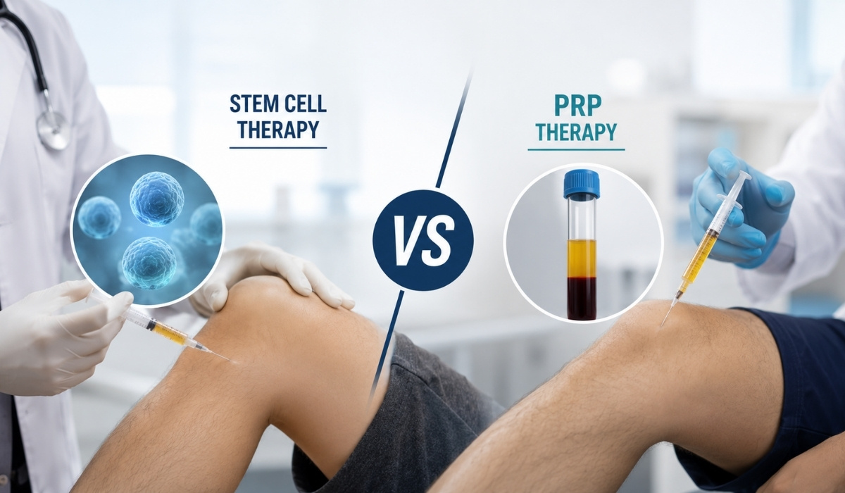
Stem Cell Therapy vs PRP Therapy comparison for knee pain treatment showing regenerative medicine injection procedures

Quick Answer

Neither treatment is universally “better” — the right choice depends on how advanced your condition is. PRP is generally the first-line regenerative option for mild-to-moderate joint pain and tendon injuries, offering good evidence, lower cost, and minimal downtime. Stem cell therapy is typically reserved for more advanced joint degeneration or cases where PRP hasn’t given lasting relief, and comes with a higher cost and a less mature (though growing) evidence base. Many patients do well starting with PRP and stepping up to stem cell therapy only if needed.


Quick Summary

  • PRP uses your own concentrated platelets; stem cell therapy uses regenerative cells capable of differentiating into tissue-specific cells.
  • PRP has stronger, more consistent clinical evidence for mild-to-moderate osteoarthritis and tendon injuries.
  • Stem cell therapy shows promise for moderate-to-severe joint degeneration, but evidence is still evolving.
  • PRP is less invasive, cheaper, and has a shorter recovery window than stem cell therapy.
  • Stem cell therapy generally costs more and may need advanced evaluation before treatment.
  • Neither treatment cures arthritis or reverses joint damage — both aim to relieve pain and support function.
  • The right choice depends on your diagnosis, severity, prior treatments, and goals — not marketing claims.
  • A qualified interventional pain specialist should assess imaging and history before recommending either option.

Introduction

If you’ve been living with joint pain, a stubborn tendon injury, or early-stage osteoarthritis, chances are you’ve come across two terms again and again while researching non-surgical options: PRP (platelet-rich plasma) therapy and stem cell therapy. Both fall under the umbrella of regenerative medicine, both use substances derived from your own body, and both are marketed as alternatives to surgery. That overlap is exactly why so many patients get confused about which one actually fits their situation.

At Nexus Advanced Pain Management in Ahmedabad, this is one of the most common questions we hear from patients exploring non-surgical pain treatment in Ahmedabad. This article breaks down how PRP and stem cell therapy actually work, where each one shines, what the evidence says, and how to think about cost and recovery — so you can have an informed conversation with your pain specialist rather than choosing based on which clinic markets harder.

What Is PRP Therapy?

Platelet-rich plasma (PRP) therapy involves drawing a small sample of your own blood, spinning it in a centrifuge to concentrate the platelets, and injecting that concentrate into the injured or painful area. Platelets release growth factors that signal the body to accelerate its own natural healing and reduce inflammation at the injection site.

  • Best suited for: tendon injuries, ligament strains, mild-to-moderate osteoarthritis, and early cartilage wear.
  • Procedure type: minimally invasive, typically completed in under an hour.
  • Recovery: most patients resume normal activity within a few days.

What Is Stem Cell Therapy?

Stem cell therapy uses cells capable of transforming into various specialized cell types — including cartilage, bone, and connective tissue. In pain management, these cells are most often harvested from the patient’s own bone marrow or adipose (fat) tissue, then processed and injected into the damaged joint or tissue with the goal of supporting tissue repair rather than just reducing inflammation.

  • Best suited for: moderate-to-severe joint degeneration and cases where PRP alone hasn’t provided lasting relief.
  • Procedure type: more involved, often requiring a harvesting step (bone marrow or fat aspiration) before injection.
  • Recovery: generally longer than PRP, with more variability between patients.

Key Differences Between PRP and Stem Cell Therapy

FactorPRP TherapyStem Cell Therapy
Source MaterialConcentrated platelets from your bloodRegenerative cells from bone marrow or fat tissue
MechanismBoosts the body’s natural healing responseSupports tissue repair and regeneration directly
Best ForMild-to-moderate OA, tendon/ligament injuriesModerate-to-severe joint degeneration
InvasivenessLow — single blood draw and injectionModerate — harvesting step plus injection
Evidence BaseStronger, more consistent across studiesGrowing, but still less mature
Typical Cost (India)LowerHigher
DowntimeMinimal, daysSlightly longer, individualized
Number of SessionsOften 1–3Usually 1, occasionally repeated

What Does the Evidence Say?

Clinical research on both treatments has expanded considerably in recent years. For mild-to-moderate knee osteoarthritis and tendon conditions, multiple studies and meta-analyses show PRP outperforming older injection options like corticosteroids for pain relief and function, with a favorable safety profile. Stem cell therapy has a smaller but growing body of evidence, with several trials reporting meaningful improvement in pain and function scores for patients with moderate-to-advanced osteoarthritis — particularly those who haven’t responded well to PRP or hyaluronic acid.

It’s worth being clear-eyed here: neither treatment reverses arthritis or guarantees a specific outcome. Response varies from patient to patient based on the severity of joint damage, overall health, and how the treatment is administered. Any clinic that promises a guaranteed cure with either treatment is not giving you an evidence-based picture.

Expert Insight

“In our experience, the biggest factor in outcome isn’t which treatment you pick — it’s how accurately the underlying condition has been diagnosed. A patient with early cartilage wear treated with PRP often does just as well as one who jumps straight to stem cell therapy, at a fraction of the cost.” — Nexus Pain Clinical Team


Safety and Risk Considerations

Both treatments use material derived from your own body, which significantly lowers the risk of allergic reaction or rejection compared to synthetic injectables. That said, both carry standard injection-related risks: temporary soreness, swelling, or infection at the injection site. Stem cell therapy, because it involves a harvesting step, carries a slightly broader risk profile simply due to the additional procedure involved.

  • Discuss your full medical history, including blood thinners or clotting conditions, before either procedure.
  • Ask whether the clinic uses image guidance (ultrasound or fluoroscopy) for injection accuracy.
  • Clarify how the platelet or cell concentration is processed and verified before injection.

Cost Comparison: PRP vs Stem Cell Therapy

Cost is one of the most common deciding factors for patients in Ahmedabad and across Gujarat. As a general pattern, PRP therapy costs meaningfully less than stem cell therapy per session, largely because the preparation process is simpler and doesn’t require a separate harvesting procedure. Stem cell therapy’s higher cost reflects the additional processing, equipment, and clinical time involved. Exact pricing varies by clinic, the joint or area being treated, and the number of sessions recommended — your pain specialist can provide a personalized estimate after evaluation.

Common Mistakes to Avoid

1. Choosing stem cell therapy over PRP simply because it sounds more “advanced,” without a clinical reason.

2. Expecting a single injection to permanently reverse years of joint wear.

3. Skipping proper imaging or diagnosis and going straight to a regenerative treatment based on symptoms alone.


Myths vs Facts

MythFact
Stem cell therapy always works better than PRP.Effectiveness depends on the condition and its severity — PRP outperforms in many mild-to-moderate cases.
PRP and stem cell therapy are the same thing.They use different biological material and work through different mechanisms.
These treatments can cure arthritis.Both aim to relieve pain and improve function; neither reverses underlying joint damage.
More expensive automatically means more effective.Cost reflects the complexity of preparation, not a guarantee of better outcomes for every patient.

Questions Patients Forget to Ask

  • How many years of experience does the specialist have performing this specific procedure?
  • Is image guidance used to ensure precise injection placement?
  • What realistic percentage of patients with my condition see meaningful improvement?
  • What is the plan if this treatment doesn’t provide sufficient relief?
  • How is the platelet or cell sample processed, and is it done in-house?

Practical Action Plan

  1. Get an accurate diagnosis with clinical examination and relevant imaging (X-ray/MRI as needed).
  2. Discuss severity grading — mild-to-moderate cases often start with PRP.
  3. Ask about evidence specific to your condition, not general marketing claims.
  4. Compare cost and expected number of sessions for both options with your specialist.
  5. Set realistic expectations — track pain and function over 6–12 weeks post-treatment.

Nexus Pain Expert Summary

PRP and stem cell therapy are both valuable tools in non-surgical pain management, but they aren’t interchangeable. PRP is typically the appropriate first step for mild-to-moderate joint pain and tendon injuries, offering solid evidence, lower cost, and quick recovery. Stem cell therapy becomes more relevant for advanced degeneration or when PRP hasn’t delivered lasting results. The most important step isn’t picking a treatment name — it’s getting an accurate diagnosis from a qualified interventional pain specialist who can match the treatment to your specific condition.

Key Takeaways

  • PRP and stem cell therapy work differently — one boosts natural healing, the other supports tissue regeneration directly.
  • PRP generally suits mild-to-moderate conditions; stem cell therapy suits more advanced degeneration.
  • PRP is less invasive and more affordable; stem cell therapy costs more due to added processing.
  • Neither treatment guarantees a cure — both aim for meaningful pain relief and improved function.
  • An accurate diagnosis matters more than which treatment name sounds more advanced.
  • Always ask about evidence, technique (image guidance), and realistic outcomes before deciding.

How Nexus Pain Can Help

Choosing between PRP and stem cell therapy starts with an accurate, individualized assessment — not a one-size-fits-all recommendation. At Nexus Advanced Pain Management, patients receive a comprehensive pain assessment that considers imaging, severity, and prior treatment history before any regenerative option is discussed.

  • Comprehensive pain assessment and diagnostic evaluation
  • Evidence-based treatment planning tailored to condition severity
  • Access to non-surgical pain management and interventional pain procedures
  • Personalized treatment planning rather than a default protocol

FAQ Section

Is stem cell therapy better than PRP for knee pain?

Not universally. PRP tends to work well for mild-to-moderate knee osteoarthritis, while stem cell therapy is generally considered for more advanced degeneration or when PRP hasn’t provided lasting relief.

What is the main difference between PRP and stem cell therapy?

PRP uses concentrated platelets from your blood to boost natural healing, while stem cell therapy uses regenerative cells from bone marrow or fat tissue that can support direct tissue repair.

Which treatment is more affordable, PRP or stem cell therapy?

PRP is generally less expensive because its preparation process is simpler and doesn’t require a separate harvesting procedure like stem cell therapy does.

How long does pain relief from PRP last?

Relief duration varies by patient and condition, but many patients report benefits lasting several months to about a year, particularly for tendon injuries and mild-to-moderate osteoarthritis.

How long does pain relief from stem cell therapy last?

Duration varies widely between patients and depends on the severity of joint damage; some patients report benefits lasting a year or more, though more research is still needed.

Is stem cell therapy safe?

Since the cells are typically taken from your own body, the risk of rejection is low. As with any injection-based procedure, there are standard risks like soreness or infection at the injection site.

Can PRP and stem cell therapy be combined?

In some cases, specialists may consider combining approaches depending on the condition, though this decision should always be based on individual evaluation rather than a default protocol.

Do PRP or stem cell injections hurt?

Most patients report mild discomfort similar to a routine injection. Local anesthesia and image guidance are often used to minimize discomfort and improve accuracy.

How many sessions of PRP or stem cell therapy are needed?

PRP is often given in one to three sessions spaced a few weeks apart, while stem cell therapy is more commonly a single session, occasionally repeated based on response.

Are PRP and stem cell therapy covered by insurance in India?

Coverage varies significantly by insurer and policy. It’s best to confirm directly with your insurance provider and the treating clinic before proceeding.

Who is not a good candidate for these treatments?

Patients with active infections, certain blood disorders, or very advanced bone-on-bone joint damage may not be ideal candidates — a clinical evaluation determines suitability.

Where can I get PRP or stem cell therapy in Ahmedabad?

Nexus Advanced Pain Management in Ahmedabad offers evaluation and evidence-based regenerative treatment planning for eligible patients as part of its non-surgical pain management services.