Categories Neck Pain

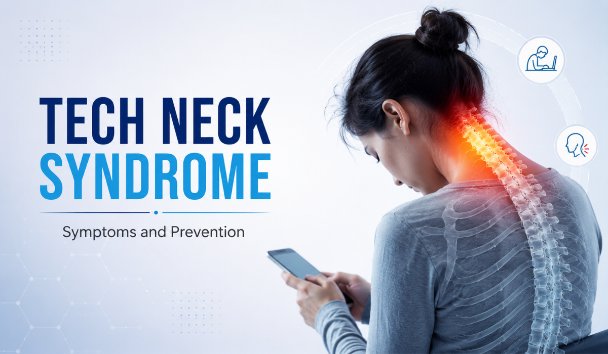
Tech Neck Syndrome: Symptoms and Prevention

tech neck syndrome

Quick Answer

 Tech neck syndrome is neck and upper-back strain caused by prolonged forward head posture while looking down at phones, laptops, or tablets. Common symptoms include neck stiffness, upper back pain, headaches, and rounded shoulders. It is generally manageable with posture correction, ergonomic changes, targeted exercises, and — when pain persists — evaluation by a pain management specialist.

Quick Summary

Key Takeaways at a Glance

●        Tech neck results from repeated forward head tilt while using digital devices.

●        Every inch the head tips forward multiplies the load on the cervical spine.

●        Common symptoms: neck stiffness, headaches, shoulder tightness, and reduced mobility.

●        Desk workers, students, and frequent smartphone users are at highest risk.

●        Most cases improve with posture correction, ergonomic adjustments, and physiotherapy.

●        Ignoring symptoms for years can contribute to long-term postural and spinal strain.

●        Persistent or worsening pain should be evaluated by a pain management specialist.

Introduction

If your neck feels stiff by mid-afternoon, or you reach for a phone and instantly feel a pull at the base of your skull, you are not alone. “Tech neck” has become one of the most common postural complaints seen in clinics today, driven by hours spent looking down at smartphones, laptops, and tablets. It is not a formal medical diagnosis in itself, but a recognizable pattern of strain that, over time, can affect the muscles, joints, and nerves of the cervical spine.

This article explains what tech neck syndrome is, why it develops, how to recognize it, and — most importantly — practical, evidence-based ways to prevent and manage it.

What Is Tech Neck Syndrome?

Tech neck syndrome, sometimes called “text neck,” describes the cumulative strain placed on the neck and upper back from repeatedly holding the head in a forward, tilted position while using digital devices. The human head weighs roughly 4.5–5.5 kg in a neutral position, but as it tips forward, the effective load on the cervical spine increases sharply — tilting to just 60 degrees can place the equivalent of 27 kg of force through the neck muscles and joints.

Over months or years, this repeated loading can lead to muscle fatigue, joint irritation, and postural changes such as forward head posture and rounded shoulders.

Symptoms of Tech Neck Syndrome

  • Persistent stiffness or tightness at the base of the neck
  • Dull, aching pain across the upper back and shoulders
  • Tension-type headaches, often starting at the back of the head
  • Reduced range of motion when turning or tilting the head
  • Rounded shoulders or a visibly forward-jutting head posture
  • Tingling or numbness radiating into the arms in more advanced cases
  • Fatigue in the neck and shoulder muscles by the end of the day

Symptoms are usually mild and intermittent at first, which is why many people delay addressing them until the discomfort becomes a daily occurrence.

Causes of Tech Neck Syndrome

  • Prolonged smartphone use with the head tilted downward
  • Laptop use without an external monitor, keyboard, or stand
  • Poor desk ergonomics — screens positioned too low or too far
  • Long, uninterrupted screen sessions without posture breaks
  • Weak deep neck flexor and upper back muscles
  • Sleeping positions that keep the neck flexed or unsupported

Risk Factors

  • Desk-based and remote workers spending 6+ hours on screens daily
  • Students using tablets or phones for extended study sessions
  • Individuals with pre-existing postural imbalances
  • People who primarily use smartphones rather than larger screens
  • Sedentary lifestyle with limited upper back and core strengthening

How Tech Neck Is Diagnosed

Tech neck is typically identified through a clinical assessment rather than imaging alone. A pain specialist or physiotherapist will usually review posture, screen-use habits, muscle tenderness, and neck range of motion. If symptoms are more advanced, persistent, or accompanied by arm numbness, imaging such as an X-ray or MRI may be recommended to rule out underlying cervical spine conditions like disc degeneration or nerve compression.

Treatment Options

Non-Surgical Treatment Approaches

  • Postural retraining and ergonomic correction
  • Targeted physiotherapy to strengthen deep neck flexors and upper back muscles
  • Stretching routines for the chest, upper trapezius, and levator scapulae
  • Heat therapy or therapeutic massage for muscle tension
  • Activity modification — scheduled screen breaks and device positioning

Most people with tech neck respond well to conservative, non-surgical care, and surgery is rarely required. If imaging and clinical evaluation confirm an underlying structural cervical condition rather than simple postural strain, a pain specialist may discuss evidence-based interventional options such as radiofrequency ablation or a targeted nerve block — but these are not first-line treatments for tech neck itself.

Prevention Tips

  • Hold devices at eye level rather than looking down
  • Position laptop or monitor screens at eye height using a stand
  • Follow the 20-20-20 rule — every 20 minutes, look 20 feet away for 20 seconds
  • Take a posture or movement break every 30–45 minutes
  • Strengthen upper back and neck-supporting muscles regularly
  • Use a supportive pillow that keeps the neck in a neutral position while sleeping
  • Set up a proper ergonomic workstation, even when working from home

Recovery and Rehabilitation

Recovery timelines vary depending on how long symptoms have been present. Mild, recently developed tech neck often improves within 2–4 weeks of consistent posture correction and stretching. More established cases with muscle imbalance may take 6–12 weeks of structured physiotherapy to see meaningful, lasting improvement. Long-term prevention depends on sustaining ergonomic habits even after symptoms resolve, since the underlying screen-use patterns typically remain unchanged.

Tech Neck vs. Cervical Spondylosis

FeatureTech Neck SyndromeCervical Spondylosis
Primary CauseProlonged forward head posture, muscular strainAge-related degeneration of cervical discs and joints
Typical Age GroupCommon across working-age adults and studentsMore common after age 40–45
Structural ChangesUsually none on imaging in early stagesDisc thinning, bone spurs visible on imaging
ReversibilityOften improves fully with posture and exerciseManageable, but degenerative changes are not reversible
Typical TreatmentErgonomics, physiotherapy, stretchingPhysiotherapy, pain management, occasionally interventional care

Expert Insight Sections

Expert Insight #1 — The 60-Degree Problem

A common mistake is assuming neck pain only comes from awkward sitting. In reality, even a mild 45–60 degree forward head tilt — the average angle used while texting — multiplies spinal load several times over. Correcting device height is often more impactful than correcting the chair.

Expert Insight #2 — Muscle Imbalance Is the Real Driver

Tech neck is rarely just about the neck. Tight chest muscles paired with weak upper back and deep neck flexor muscles create the classic “upper crossed” pattern. Long-term relief usually requires strengthening the weak muscles, not just stretching the tight ones.

Expert Insight #3 — When to Stop Self-Managing

If symptoms persist beyond 4–6 weeks of consistent posture and exercise changes, or if numbness or tingling develops in the arms, this is a signal to see a pain specialist rather than continuing to self-treat.

Common Mistakes

  1. Only stretching the tight muscles while ignoring the weak, underactive ones that need strengthening.
  2. Raising the phone briefly for photos but reverting to a downward gaze during normal browsing and messaging.
  3. Waiting until pain becomes constant before seeking a professional assessment, allowing postural patterns to become deeply ingrained.

Myths vs. Facts

MythFact
Tech neck only affects people who work at a desk all day.Frequent smartphone use alone — even without a desk job — can cause tech neck.
Tech neck always requires surgery to fix.The large majority of cases improve with posture correction and physiotherapy; surgery is rarely needed.
A quick stretch once a day is enough to reverse it.Consistent, structured strengthening over several weeks is usually needed for lasting change.
Neck pain from screens will simply go away on its own.Without addressing posture and screen habits, symptoms often persist or gradually worsen.

Questions Patients Forget to Ask

  • Could my headaches be related to my neck posture rather than a separate issue?
  • Should I get an X-ray or MRI, or is a clinical exam sufficient at this stage?
  • What specific exercises should I be doing, and how often?
  • Is my sleeping position contributing to the problem?
  • How will I know if my tech neck is improving versus progressing?

Practical Action Plan

  1. This week: Raise your phone and laptop screen to eye level and set a 30-minute break reminder.
  2. This week: Begin gentle chin-tuck and shoulder blade squeeze exercises, 2–3 sets daily.
  3. Next 2–4 weeks: Track whether stiffness and headaches are reducing in frequency.
  4. If no improvement after 4 weeks: Book an evaluation with a pain management or physiotherapy specialist.
  5. Ongoing: Reassess your workstation ergonomics every few months, especially after any equipment changes.

Nexus Pain Expert Summary

Tech neck syndrome is a manageable, posture-driven condition in the vast majority of cases. Early recognition and consistent ergonomic and exercise habits prevent most cases from progressing. Persistent pain, recurring headaches, or arm symptoms warrant a proper clinical evaluation to rule out underlying cervical spine involvement and to build a personalized, non-surgical management plan.

How Nexus Pain Can Help

For patients in Ahmedabad experiencing persistent neck stiffness, headaches, or postural discomfort linked to screen use, Nexus Advanced Pain Management offers a structured, evidence-based approach to assessment and care, including:

  • Comprehensive pain and postural assessment
  • Evidence-based, personalized treatment planning
  • Non-surgical pain management strategies
  • Guidance on ergonomic and lifestyle modification
  • Rehabilitation support for long-term postural correction
  • Long-term strategies to prevent symptom recurrence

The goal of care is not just short-term symptom relief but building sustainable habits that address the root cause of tech neck.

Key Takeaways

  • Tech neck stems from repeated forward head posture during screen use.
  • Device height and screen breaks matter more than most people realize.
  • Strengthening — not just stretching — is key to lasting relief.
  • Most cases resolve without surgery through posture and physiotherapy-based care.
  • Persistent symptoms beyond 4–6 weeks should be professionally assessed.
  • Sleep posture and workstation setup are frequently overlooked contributors.

FAQ Section

1. What is tech neck syndrome?

Tech neck syndrome is neck and upper back strain caused by prolonged forward head posture while using phones, laptops, or tablets.

2. What are the first signs of tech neck?

Early signs include mild neck stiffness, tightness at the base of the skull, and tension headaches, especially after long screen sessions.

3. Can tech neck cause headaches?

Yes. Muscle tension at the base of the skull from forward head posture commonly triggers tension-type headaches.

4. Is tech neck permanent?

In most cases, tech neck is not permanent and improves significantly with posture correction, ergonomic changes, and targeted exercise.

5. How long does it take to fix tech neck?

Mild cases often improve within 2–4 weeks; more established postural patterns may take 6–12 weeks of consistent care.

6. Can children and teenagers get tech neck?

Yes, students who use tablets and phones for long study or gaming sessions are increasingly seen with early tech neck symptoms.

7. Does tech neck cause arm numbness?

In more advanced cases, nerve irritation from prolonged strain can cause tingling or numbness radiating into the arms, which warrants specialist evaluation.

8. What is the best sleeping position to avoid tech neck?

Sleeping on your back or side with a supportive pillow that keeps the neck in a neutral, aligned position is generally best.

9. Is tech neck the same as cervical spondylosis?

No. Tech neck is a posture-driven muscular strain pattern, while cervical spondylosis involves age-related degenerative changes to the cervical spine, visible on imaging.

10. What exercises help with tech neck?

Chin tucks, shoulder blade squeezes, upper back strengthening, and chest stretches are commonly recommended starting exercises.

11. When should I see a pain specialist for tech neck?

If symptoms persist beyond 4–6 weeks despite posture correction, or if you notice arm numbness, weakness, or worsening headaches, a specialist evaluation is advised.

12. Can tech neck be prevented completely?

While occasional device use is unavoidable, consistent ergonomic habits and regular movement breaks significantly reduce the risk of developing symptoms.


Categories Back Pain

Best Sleeping Positions for Back Pain Relief

Best Sleeping Poistions for Back Pain Relief

Quick Answer

• The best sleeping positions for back pain relief are side sleeping with a pillow between the knees, or back sleeping with a pillow under the knees.

• Both positions keep the spine in a neutral, aligned position and reduce pressure on the lower back.

• Stomach sleeping is generally the least supportive position and can strain the neck and lower spine.

Quick Summary

  • Side sleeping with a pillow between the knees keeps the hips, pelvis, and spine aligned.
  • Back sleeping with a small pillow under the knees reduces pressure on the lumbar spine.
  • Stomach sleeping is the least recommended position for most people with back pain.
  • Pillow height and mattress firmness matter as much as the position itself.
  • The fetal position can help open the space between vertebrae for people with a herniated disc.
  • It typically takes one to two weeks of consistent practice to adjust to a new sleep position.
  • Morning back pain that does not ease with position changes may need a professional evaluation.

Introduction

Most people spend close to a third of their life asleep, yet very few think about how their sleeping position affects their spine. For someone already managing back pain, the wrong posture at night can undo an entire day of careful movement, stretching, and rest. The good news is that sleep position is one of the simplest and most controllable factors in reducing back pain, and adjusting it does not require expensive equipment or medical intervention in most cases.

This guide walks through the sleeping positions that are best for back pain relief, the ones to avoid, why sleep posture matters, and when ongoing back pain during sleep is a sign that it is time to speak with a pain management specialist.

How Sleep Position Affects Back Pain

The spine has natural curves at the neck, mid-back, and lower back. When a person lies down in a position that does not support these curves, muscles around the spine work overtime to compensate, and the discs and joints between vertebrae come under uneven pressure. Over an eight-hour night, this uneven loading can leave the lower back stiff, sore, or noticeably worse by morning.

A supportive sleep position does the opposite: it keeps the spine close to its natural, neutral alignment, allows the muscles around it to relax fully, and reduces the load placed on the discs and facet joints. This is why the right combination of position, pillow placement, and mattress support often brings a noticeable improvement within one to two weeks.

The Best Sleeping Positions for Back Pain Relief

1. Side Sleeping With a Pillow Between the Knees

Lying on the side with the knees slightly bent and a firm pillow placed between them is one of the most consistently recommended positions for lower back pain. The pillow keeps the top leg from pulling the pelvis and lower spine out of alignment, which reduces strain on the lower back and hips through the night.

2. Back Sleeping With a Pillow Under the Knees

Sleeping on the back with a small pillow or rolled towel placed under the knees helps maintain the natural curve of the lower spine and takes pressure off the lumbar discs. This position distributes body weight evenly across the widest surface of the body, which many people with general lower back stiffness find comfortable.

3. Fetal Position for Disc-Related Pain

For people with a herniated or bulging disc, curling gently onto one side with the knees drawn toward the chest can open up the space between vertebrae and ease pressure on a compressed nerve. Switching sides periodically through the night helps avoid one-sided muscle tightness.

4. Slightly Reclined Position for Certain Conditions

For some patients, particularly those recovering from spinal surgery or managing spinal stenosis, sleeping in a slightly reclined position, such as in an adjustable bed or with extra pillows under the upper back, can reduce pressure on the lower spine. This position should generally be guided by a treating specialist rather than adopted on its own.

Sleeping Position to Avoid: Stomach Sleeping

Stomach sleeping is the position most likely to aggravate back and neck pain. It flattens the natural curve of the lower spine and forces the neck to stay turned to one side for hours, which strains both the cervical and lumbar spine. Anyone who currently sleeps on their stomach and struggles to change positions can place a thin pillow under the pelvis and lower abdomen to reduce the arch in the lower back while gradually transitioning to side or back sleeping.

Symptoms That Suggest Sleep Position Is Contributing to Back Pain

  • Back pain that feels worse first thing in the morning and eases within an hour of moving around.
  • Stiffness concentrated on one side of the lower back or hip.
  • Tingling or numbness in the legs that appears after lying in one position for a long time.
  • Pain that improves noticeably on nights spent away from a usual bed, such as while travelling.

Causes and Risk Factors

Sleep-related back pain is rarely caused by position alone. It usually develops from a combination of factors that make the spine more sensitive to how it is loaded overnight.

  • An unsupportive or sagging mattress that no longer holds the spine in alignment.
  • Long hours of sitting during the day that leave hip and back muscles tight before bed.
  • Existing conditions such as a herniated disc, sciatica, spinal stenosis, or facet joint arthritis.
  • Excess strain from stomach sleeping or sleeping without any knee or lumbar support.
  • Weak core and back muscles that are less able to stabilise the spine during sleep.

When to See a Specialist

Adjusting sleep position and pillow support resolves mild, mechanical back pain for many people. However, certain signs suggest the pain needs a proper clinical evaluation rather than home adjustments alone.

  • Pain that radiates down one or both legs, especially with numbness or tingling.
  • Back pain that wakes you from sleep or is unrelieved by any position.
  • Pain lasting more than four to six weeks despite changes in sleep posture and mattress support.
  • Any loss of bladder or bowel control, which requires immediate medical attention.

A pain management specialist can assess whether the pain is coming from a disc, joint, nerve, or muscular source, and confirm whether a change in sleep habits alone is enough or whether further treatment is needed.

Non-Surgical Treatment Options

For most patients, back pain linked to sleep posture and general spinal strain responds well to non-surgical, evidence-based care rather than surgery. Options a pain management team may consider include:

  • Targeted physiotherapy and core-strengthening exercises to improve spinal support.
  • Postural correction and ergonomic guidance for both daytime and sleep positions.
  • Interventional options such as epidural injections or nerve blocks for nerve-related pain.
  • Radiofrequency ablation for chronic facet joint pain that has not responded to conservative care.
  • Structured lifestyle and weight management guidance where it is contributing to spinal load.

Prevention Tips for Better Sleep Posture

  • Choose a medium-firm mattress that supports the spine without being too soft or too hard.
  • Replace pillows that have flattened or lost their shape, typically every twelve to eighteen months.
  • Keep a consistent sleep position for most of the night rather than shifting constantly.
  • Do a few minutes of gentle stretching before bed to release tension in the hips and lower back.
  • Avoid falling asleep on the couch in a slouched or twisted position.

Recovery and Adjustment Period

Switching to a more supportive sleep position rarely feels comfortable on the first night. Most people need one to two weeks of consistent practice, along with correctly placed pillows, before the new position feels natural. Setting reminders to check position before falling asleep, and using a body pillow to prevent rolling onto the stomach, can make the transition easier. If pain increases rather than eases after two to three weeks of a genuine, consistent change, it is a reasonable point to seek a professional opinion.

Comparison of Sleeping Positions for Back Pain

PositionEffect on SpineBest ForWatch-Outs
Side with pillow between kneesKeeps hips and spine alignedGeneral lower back pain, pregnancy-related painUse a firm enough pillow to fill the gap between knees
Back with pillow under kneesMaintains natural lumbar curveGeneral stiffness, even weight distributionNot ideal for those with sleep apnoea or snoring
Fetal positionOpens space between vertebraeHerniated or bulging disc painAlternate sides to avoid one-sided tightness
Slightly reclinedReduces lumbar loadPost-surgical recovery, spinal stenosisBest used under specialist guidance
Stomach sleepingFlattens spinal curve, strains neckGenerally not recommendedTransition out gradually rather than all at once

Expert Insight

Expert Insight 1: Pillow Placement Matters as Much as Position

• A side sleeper without a knee pillow can still let the top leg roll forward, twisting the lower spine even in an otherwise good position.

• Practical takeaway: place the pillow high enough between the knees that the hips stay level.


Expert Insight 2: Mattress Age Is an Overlooked Factor

• A mattress that has sagged in the middle can undo the benefit of an otherwise correct sleep position.

• Practical takeaway: if a mattress is more than seven to eight years old and shows visible sagging, it is worth having it assessed or replaced.


Expert Insight 3: One Bad Night Is Not a Diagnosis

• Occasional stiffness after an unusual sleeping arrangement, such as travel, is common and not usually a sign of a serious problem.

• Practical takeaway: track whether the pain is a one-off or a pattern over one to two weeks before assuming a deeper issue.

Common Mistakes

  1. Using a pillow that is too soft to keep the knees, hips, and spine properly separated and aligned.
  2. Trying to force a completely new sleep position overnight instead of transitioning gradually.
  3. Ignoring persistent morning pain for months instead of seeking an evaluation after four to six weeks.

Myths vs Facts

MythFact
A firmer mattress is always better for back pain.A medium-firm mattress that supports the spine’s natural curve is generally more effective than an extremely firm one.
Back sleeping is bad for everyone with back pain.Back sleeping with knee support works well for many people; it depends on the underlying cause of the pain.
Sleeping position alone can fix any back pain.Position helps with mechanical, posture-related pain but does not replace treatment for disc, nerve, or joint conditions.
You should sleep in whatever position feels easiest to fall asleep in.The easiest position to fall asleep in is not always the one that protects the spine through the whole night.

Questions Patients Forget to Ask

  • Could my mattress or pillow be contributing to my pain, not just my sleep position?
  • Is my pain more likely to be coming from a disc, a joint, or a muscle?
  • Are there specific stretches I should do before bed for my particular type of back pain?
  • At what point should I stop trying home adjustments and come in for an evaluation?

Practical Action Plan

  1. Identify your current default sleep position and note where morning pain is concentrated.
  2. Switch to side sleeping with a knee pillow or back sleeping with a pillow under the knees.
  3. Check your mattress and pillow for sagging or flattening, and replace if needed.
  4. Track your pain for two weeks after the change to see if it is improving, unchanged, or worsening.
  5. If pain persists, radiates, or disrupts sleep after this period, book an evaluation with a pain management specialist.

Nexus Pain Expert Summary 

• Side sleeping with a knee pillow or back sleeping with a pillow under the knees are the most supportive positions for most people with back pain.

• Stomach sleeping should generally be avoided or transitioned out of gradually.

• Give a new sleep position one to two weeks before judging its effect, and seek a professional evaluation if pain persists, radiates, or disrupts sleep.

How Nexus Pain Can Help

For back pain that does not improve with sleep posture changes and basic home care, Nexus Pain Management offers a structured, evidence-based approach. This includes a comprehensive pain assessment to identify whether the source is a disc, joint, nerve, or muscular in nature, followed by a personalised, non-surgical treatment plan. Depending on the assessment, this may involve targeted rehabilitation guidance, interventional pain procedures, or long-term pain management strategies designed around each patient’s daily routine, including practical, sustainable lifestyle and sleep posture modifications.

Key Takeaways

  • Sleep posture directly affects spinal alignment and how rested the back feels each morning.
  • Side sleeping with a knee pillow and back sleeping with a knee pillow are the two most supportive options.
  • Stomach sleeping places the most strain on the neck and lower back.
  • Mattress and pillow condition matter as much as the position itself.
  • Give any new sleep position one to two weeks before evaluating its effect.
  • Radiating pain, numbness, or pain lasting beyond four to six weeks warrants a specialist evaluation.

Frequently Asked Questions

  1. What is the best sleeping position for back pain?

For most people, side sleeping with a pillow between the knees or back sleeping with a pillow under the knees provides the most spinal support.

  1. Is it bad to sleep on your stomach if you have back pain?

Stomach sleeping generally flattens the lower spine’s natural curve and strains the neck, so it is usually the least recommended position for back pain.

  1. How long does it take to adjust to a new sleeping position?

Most people need about one to two weeks of consistent practice before a new sleep position feels natural and comfortable.

  1. What is the best sleeping position for sciatica?

Many people with sciatica find relief in the fetal position on the side that is not affected, or back sleeping with a pillow under the knees.

  1. Should I sleep on a firm or soft mattress for back pain?

A medium-firm mattress that supports the spine’s natural curve is generally recommended over one that is extremely soft or extremely hard.

  1. Can a pillow really make a difference to back pain?

Yes. A pillow placed between the knees or under the knees helps keep the hips and spine aligned, which reduces strain through the night.

  1. Why does my back hurt more in the morning than during the day?

This often happens when a sleep position places uneven pressure on the spine for several hours, and it usually eases once movement resumes.

  1. Is it normal for back pain to worsen while traveling or sleeping away from home?

Yes, an unfamiliar mattress or pillow can temporarily disrupt spinal support, though this is usually short-lived and not a sign of a new problem.

  1. Can sleeping position alone cure chronic back pain?

Sleeping position can meaningfully reduce mechanical, posture-related pain, but underlying conditions such as a disc or nerve issue usually need a broader treatment plan.

  1. When should I see a pain specialist about back pain during sleep?

If pain radiates down a leg, includes numbness, wakes you from sleep, or lasts beyond four to six weeks despite position changes, it is time for an evaluation.

  1. Are adjustable beds helpful for back pain?

A slightly reclined position can help some patients, particularly those recovering from surgery or managing spinal stenosis, but this is best guided by a specialist.

  1. Where can I get a professional evaluation for back pain in Ahmedabad?

Nexus Pain Management in Ahmedabad offers comprehensive assessments and evidence-based, non-surgical treatment plans for back pain that does not improve with home adjustments.


Categories Knee Pain

Why Do Knees Hurt While Climbing Stairs?

Quick Answer

Knees hurt while climbing stairs because stairs place 3–4 times your body weight through the kneecap joint, far more than walking on flat ground. Pain is most often caused by patellofemoral pain syndrome (irritation under the kneecap), early cartilage wear, weak thigh muscles, or, in older adults, osteoarthritis. Most cases improve with targeted strengthening, activity modification, and non-surgical pain management — surgery is rarely the first step.

Quick Summary

  • Stairs load the kneecap joint 3–4x more than flat-ground walking, which is why pain often appears only on stairs.
  • Descending is usually more painful than climbing because it requires the thigh muscles to control your body weight while lengthening (eccentric load).
  • The most common cause in adults under 45 is patellofemoral pain syndrome; in adults over 50, early knee osteoarthritis is more likely.
  • Weak or imbalanced thigh (quadriceps) muscles are a leading, correctable contributor.
  • Pain that lasts more than 2–3 weeks, causes swelling, or is accompanied by locking or giving-way needs clinical evaluation.
  • Most stair-related knee pain responds well to physiotherapy, activity modification, and non-surgical interventional treatment.
  • A pain management specialist can identify the exact source of pain using clinical assessment and imaging, avoiding unnecessary surgery.

Introduction

Climbing or descending a flight of stairs is one of the most physically demanding everyday movements for the knee joint — more demanding, in fact, than walking or even jogging on level ground. So when a patient says, “My knees are fine most of the day, but stairs are unbearable,” that single detail is clinically meaningful. It tells us the problem is very likely centered on the kneecap (patellofemoral) joint rather than the whole knee, and it usually points toward a specific, identifiable cause rather than vague “wear and tear.”

This guide explains exactly why stairs trigger knee pain, what the common underlying causes are, how pain specialists diagnose the problem, and which non-surgical treatments genuinely help. The goal is not to alarm you — the goal is to help you understand your knee well enough to make an informed decision about what to do next.

Why Stairs Are So Hard on Your Knees

Every time you climb a step, your kneecap (patella) glides within a groove at the end of the thighbone (femur). This is called the patellofemoral joint. Research in joint biomechanics has consistently shown that this joint bears roughly 3–4 times your body weight when climbing stairs, compared to about 1–1.3 times your body weight during level walking. Descending stairs can place even greater strain on the joint, because your thigh muscles must contract while lengthening to control your descent — a pattern called eccentric loading, which is more taxing on both muscle and joint surface.

Why this matters: if your knee only hurts on stairs (and not on flat ground), the problem is almost always related to how load passes through the kneecap joint — not necessarily deep, structural damage inside the knee.

Common Symptoms of Stair-Related Knee Pain

  • A dull, aching pain around or behind the kneecap when climbing or descending
  • Sharp, catching pain on the first few steps, especially going downstairs
  • A grinding, clicking, or crunching sensation (crepitus) with movement
  • Mild swelling after stair use or prolonged sitting
  • Stiffness after sitting with bent knees for a long time (“theatre sign”)
  • A feeling that the knee may “give way” on stairs, particularly descending

What Causes Knee Pain While Climbing Stairs?

1. Patellofemoral Pain Syndrome (PFPS)

The single most common cause in active adults and young patients. It occurs when the kneecap doesn’t track smoothly in its groove, causing irritation of the cartilage and surrounding tissue under repetitive loading — exactly the kind of loading stairs produce.

2. Chondromalacia Patella

A softening or early wear of the cartilage on the underside of the kneecap. It shares symptoms with PFPS but reflects an actual, visible change in the cartilage surface, usually identified on imaging.

3. Knee Osteoarthritis

In patients over 45–50, progressive cartilage thinning across the joint is a common cause. Stair pain — particularly descending — is often one of the earliest reported symptoms of osteoarthritis, appearing before pain on flat ground.

4. Quadriceps Weakness or Imbalance

The quadriceps (thigh) muscles, especially the inner portion (vastus medialis), help guide the kneecap’s movement. Weakness or imbalance here is one of the most common — and most correctable — contributors to stair pain.

5. Patellar Tendinitis (“Jumper’s Knee”)

Overuse-related inflammation of the tendon just below the kneecap, common in athletes and people who recently increased stair-climbing, running, or squatting activity.

6. Meniscus Irritation

The meniscus (shock-absorbing cartilage between the thigh and shin bones) can become irritated or, in some cases, torn, causing pain that is often worse with twisting or descending motions.

7. IT Band Syndrome

Tightness in the iliotibial band running along the outer thigh can pull on the outer kneecap structures, producing pain that is frequently worse going down stairs.

Risk Factors

  • Age over 45, particularly for osteoarthritis-related causes
  • Sedentary lifestyle with weak thigh and hip muscles
  • Sudden increase in stair use, running, or squatting activity
  • Previous knee injury or surgery
  • Being overweight, which multiplies joint loading on stairs
  • Occupations requiring frequent stair or ladder use
  • Flat feet or abnormal lower-limb alignment

How Is Stair-Related Knee Pain Diagnosed?

A pain management specialist typically begins with a detailed history — including exactly when the pain occurs (up vs. down stairs), its duration, and any swelling or instability — followed by a physical examination assessing kneecap tracking, muscle strength, and joint stability. Depending on findings, imaging such as X-ray (for joint space and arthritis) or MRI (for cartilage, tendon, or meniscus detail) may be recommended. Diagnosis is rarely guesswork; it is a structured process of ruling causes in or out.

Expert Insight

“Patients often assume stair pain automatically means arthritis, but in our clinical experience, patellofemoral pain syndrome and muscle weakness are far more common causes, particularly in patients under 50. A focused assessment usually identifies the exact source within one visit.”

Treatment Options for Knee Pain on Stairs

Non-Surgical Treatment (First-Line Approach)

For the vast majority of patients, non-surgical, evidence-based management is the appropriate starting point:

  • Targeted physiotherapy to strengthen the quadriceps, hips, and core
  • Activity and load modification (e.g., using a handrail, reducing stair frequency temporarily)
  • Anti-inflammatory strategies guided by a physician, where appropriate
  • Bracing or taping to support kneecap tracking during recovery
  • Interventional options such as targeted injections or PRP therapy for select cases of cartilage irritation or tendinitis
  • Radiofrequency ablation (RFA) for select chronic osteoarthritis-related knee pain that hasn’t responded to conservative care

Surgery is generally considered only when structural damage (such as a significant meniscus tear or advanced arthritis) is confirmed and conservative treatment has not provided adequate relief.

Prevention Tips

  • Strengthen the quadriceps and hip muscles with guided exercises 2–3 times per week
  • Warm up before activities that involve repeated stair use
  • Maintain a healthy body weight to reduce joint loading
  • Use proper footwear with adequate cushioning and support
  • Avoid sudden spikes in stair-climbing, running, or squat-based activity
  • Address flat feet or alignment issues with orthotics if recommended

Recovery and Rehabilitation

Most cases of patellofemoral pain syndrome and mild chondromalacia improve within 6–12 weeks of consistent, guided physiotherapy. Recovery is typically progressive: initial pain reduction and swelling control in the first 1–2 weeks, followed by graded strengthening over 4–8 weeks, and a gradual return to full stair activity by week 8–12. Osteoarthritis-related pain follows a longer-term management pattern rather than a fixed recovery timeline, focused on symptom control and joint preservation.

Comparison: Climbing vs. Descending Stairs

AspectClimbing (Ascending)Descending
Muscle actionConcentric (muscle shortens)Eccentric (muscle lengthens under load)
Typical joint stressHighHigher — often the more painful direction
Common associated causeQuadriceps weakness, tendinitisPFPS, chondromalacia, early osteoarthritis
Typical sensationAching, fatigue-like painSharp, catching, or unstable feeling

Common Mistakes Patients Make

  1. Ignoring stair pain for months, assuming it will “just go away”, which allows muscle weakness and altered movement patterns to worsen.
  2. Resting completely instead of modifying activity — prolonged inactivity weakens the quadriceps further and can worsen symptoms.
  3. Self-diagnosing as “arthritis” and assuming surgery is inevitable, when most cases respond well to non-surgical care.
  4. Skipping professional strengthening guidance and doing generic online exercises that may not target the right muscles.

Myths vs. Facts

MythFact
Knee pain on stairs always means arthritisIt is often patellofemoral pain syndrome or muscle weakness, especially under age 50
Rest is the best treatmentGuided activity and targeted strengthening usually work better than complete rest
Surgery is the only real fixMost patients improve significantly with non-surgical, evidence-based treatment
Clicking or cracking always signals damagePainless clicking (crepitus) is common and often not a sign of injury

Questions Patients Forget to Ask

  • Is my pain coming from the kneecap joint specifically, or the whole knee?
  • What is causing my pain — is it muscular, cartilage-related, or joint-related?
  • What is a realistic recovery timeline for my specific cause?
  • Which exercises should I avoid until my pain improves?
  • What are my non-surgical options before considering more invasive treatment?

Practical Action Plan

  1. Track your pain pattern for one week — note whether it’s worse going up, down, or both, and whether swelling occurs.
  2. Reduce (don’t eliminate) stair use temporarily and use a handrail for support.
  3. Begin gentle quadriceps and hip-strengthening exercises, ideally under guidance.
  4. Schedule an evaluation with a pain management specialist if pain persists beyond 2–3 weeks, or sooner if there is swelling, locking, or instability.
  5. Follow a structured, evidence-based treatment plan rather than a generic online routine.

Nexus Pain Expert Summary

Stair-related knee pain is common, usually explainable, and in most cases treatable without surgery. At Nexus Advanced Pain Management, we begin with a comprehensive assessment to identify the exact source of your pain, then build a personalized, non-surgical treatment plan — from targeted physiotherapy to interventional options like PRP or radiofrequency ablation where appropriate — with realistic expectations and long-term joint health as the goal.

How Nexus Pain Management Can Help

Nexus Advanced Pain Management takes an evidence-based approach to knee pain, beginning with a comprehensive assessment to identify the true source of your discomfort rather than treating symptoms generically. Our approach includes personalized, non-surgical treatment planning, interventional pain procedures where clinically appropriate, rehabilitation guidance, and long-term strategies for joint health and pain relief — all grounded in realistic expectations and patient education.

Key Takeaways

  • Stairs load the kneecap joint 3–4x more than walking, which is why pain often appears there first.
  • Descending stairs is frequently more painful than climbing due to eccentric muscle loading.
  • Common causes include patellofemoral pain syndrome, chondromalacia, quadriceps weakness, and — in older adults — early osteoarthritis.
  • Persistent pain beyond 2–3 weeks, swelling, or instability warrants professional evaluation.
  • Non-surgical treatment is effective for the large majority of patients.
  • A structured diagnosis prevents unnecessary worry and unnecessary surgery.

Frequently Asked Questions

  1. Why does my knee only hurt on stairs and not when walking?

Stairs place several times more load through the kneecap joint than flat-ground walking, so problems localized to that joint — such as patellofemoral pain syndrome — often show up only on stairs.

  1. Is it normal for knees to hurt while climbing stairs?

Occasional mild discomfort can occur with deconditioning, but persistent or worsening pain is not something to ignore and should be evaluated, particularly if it lasts more than 2–3 weeks.

  1. Why is going down the stairs more painful than going up?

Descending requires your thigh muscles to control your body weight while lengthening (eccentric loading), which places greater stress on the kneecap joint than the muscle-shortening action used while climbing.

  1. Can weak thigh muscles really cause knee pain on stairs?

Yes. Weak or imbalanced quadriceps muscles are one of the most common and most correctable contributors to stair-related knee pain, as they affect how the kneecap tracks during movement.

  1. Does knee pain on stairs mean I have arthritis?

Not necessarily. While early osteoarthritis can present this way in adults over 45–50, patellofemoral pain syndrome and muscle weakness are more common causes in younger, active patients.

  1. When should I see a pain specialist for knee pain on stairs?

See a specialist if pain persists beyond 2–3 weeks, is accompanied by swelling, clicking with pain, locking, instability, or if it is limiting your daily activities.

  1. Can knee pain on stairs be treated without surgery?

Yes. Most cases respond well to physiotherapy, activity modification, and non-surgical interventional treatments; surgery is generally reserved for confirmed structural damage that hasn’t responded to conservative care.

  1. What tests are used to diagnose stair-related knee pain?

Diagnosis typically starts with a clinical history and physical examination, followed by imaging such as X-ray or MRI when needed to assess cartilage, tendon, or joint condition.

  1. How long does it take for stair-related knee pain to improve?

Many patients with patellofemoral pain syndrome improve within 6–12 weeks of consistent, guided treatment, though timelines vary depending on the underlying cause.

  1. Can being overweight cause knee pain on stairs?

Yes. Excess body weight significantly increases the load passing through the kneecap joint during stair use, which can contribute to or worsen symptoms.

  1. Is clicking or cracking in the knee on stairs a cause for concern?

Painless clicking (crepitus) is common and usually not concerning on its own; it becomes relevant mainly when accompanied by pain, swelling, or a catching sensation.

  1. Where can I get evaluated for knee pain while climbing stairs in Ahmedabad?

Nexus Advanced Pain Management in Ahmedabad offers comprehensive knee pain assessment and evidence-based, non-surgical treatment planning for stair-related and activity-related knee pain.


Categories Botox Therapy

Botox Therapy for Migraine: How Effective Is It?

Botox therapy for migraine showing a patient receiving Botox injections for migraine relief

Quick Answer

Botox (onabotulinumtoxinA) is an evidence-backed preventive treatment specifically approved for chronic migraine — defined as 15 or more headache days a month, with migraine features on at least 8 of those days. Large clinical trials (the PREEMPT program) showed it reduces headache frequency, duration, and migraine-related disability compared to placebo when used as one of a series of quarterly injection cycles. It is not approved or well-supported for episodic migraine (fewer than 15 headache days a month), and it does not stop a migraine attack that has already started — its role is prevention, not acute relief.

Quick Summary

  • Botox for migraine is FDA-approved specifically for chronic migraine (≥15 headache days/month, with migraine features on ≥8 days).
  • The PREEMPT trials found significant reductions in headache frequency, migraine days, and disability compared to placebo over 24 weeks.
  • Treatment involves 31 small injections (155 units total) across 7 head and neck muscle areas, repeated every 12 weeks.
  • Many patients notice improvement after the second treatment cycle; a full trial usually spans 2 cycles (about 6 months) before effectiveness is assessed.
  • Common side effects are mild — neck pain, injection-site discomfort, and occasional eyelid drooping.
  • Botox is not approved for episodic migraine and is not a treatment for an active migraine attack.
  • It is typically considered after standard oral preventive medications have been tried and found insufficient.

Introduction

For patients living with frequent, disabling migraines, the idea of using Botox — a drug most associate with cosmetic wrinkle treatment — as a medical therapy can feel unfamiliar. Yet onabotulinumtoxinA is one of the few treatments specifically studied and approved for chronic migraine prevention, backed by one of the largest clinical trial programs in headache medicine. This article explains what the evidence actually shows about its effectiveness, who it’s suited for, what the treatment involves, and where it fits alongside other migraine prevention options.

What Is Chronic Migraine, and Why Does the Distinction Matter?

Chronic migraine is defined as headaches occurring on 15 or more days per month for more than three months, with migraine features present on at least 8 of those days. This is different from episodic migraine, where attacks occur less frequently. The distinction matters because Botox’s approval and the strongest evidence for its effectiveness apply specifically to chronic migraine — it has not been shown to work reliably for episodic migraine, and prescribing it outside its studied population is generally not recommended.

How Botox Works for Migraine Prevention

Botox is injected into specific muscles of the head, neck, and shoulders, where it is thought to block the release of pain-signalling chemicals and reduce activation of pain pathways involved in migraine, including peripheral nerve endings that contribute to central sensitization. Unlike acute migraine medications, which are taken during an attack, Botox works as a preventive therapy administered on a fixed schedule — it does not relieve pain once a migraine has already started.

What the Clinical Evidence Shows

The Phase III PREEMPT (Phase III REsearch Evaluating Migraine Prophylaxis Therapy) program remains the primary evidence base for Botox in chronic migraine. Across two large placebo-controlled trials involving over 1,300 patients, pooled results showed statistically significant reductions in headache-day frequency, migraine-day frequency, and headache-related disability with onabotulinumtoxinA compared to placebo. Improvements were also seen in quality-of-life measures. It’s worth noting that in the first individual trial (PREEMPT 1), the specific primary endpoint of headache-episode frequency did not reach statistical significance versus placebo, though other measures did — this is one reason the pooled analysis across both trials is considered the more complete picture of efficacy. Long-term follow-up data through multiple treatment cycles has shown continued and, in some analyses, cumulative improvement with repeated treatment.

Botox vs. Oral Preventive Medication: How It Compares

FactorBotox (OnabotulinumtoxinA)Oral Preventive Medication
Approved useChronic migraine onlyChronic and episodic migraine, depending on drug
AdministrationInjections every 12 weeks by a trained clinicianDaily or near-daily tablets
Onset of noticeable effectOften after 1–2 treatment cycles (up to ~6 months)Varies; often several weeks
Adherence burdenLow — quarterly visitsHigher — daily dosing required
Common side effectsNeck pain, injection-site discomfort, eyelid droopFatigue, weight change, mood effects (varies by drug)
Typical use caseAfter oral preventives have failed or are not toleratedUsually tried first for most patients

The Treatment Process

  • A total of 155 units of Botox is given as 31 small injections across 7 specific muscle areas of the head and neck
  • Each session takes roughly 10–15 minutes and is performed in-clinic without general anaesthesia
  • Injections are repeated approximately every 12 weeks, as the effect of each treatment cycle gradually wears off
  • Most patients require at least 2 treatment cycles (about 6 months) before their doctor can properly assess whether it’s working
  • Some patients notice initial improvement as early as 4 weeks after the first session, though this varies

Who Is a Good Candidate?

  • Adults with chronic migraine (15+ headache days a month, with migraine features on 8+ of those days) for at least 3 months
  • Patients who have not achieved adequate control with standard oral preventive medications, or who cannot tolerate them
  • Patients able to commit to quarterly injection visits and a realistic 2-cycle trial period to judge effectiveness

Botox is generally not recommended for episodic migraine, for treating an acute migraine attack in progress, or in patients with certain neuromuscular conditions or allergies to the formulation — your treating specialist will review your full history before recommending it.

Safety and Side Effects

In clinical trial data, most side effects were mild to moderate. The most commonly reported included neck pain, pain at the injection site, and temporary eyelid drooping (ptosis) or muscular weakness near the injection areas. Serious adverse events were uncommon, and discontinuation due to side effects occurred in a small minority of patients. As with any injectable treatment, it should only be administered by a trained specialist familiar with the specific migraine injection protocol.

Expert Insight

Expert Insight 1 — On Realistic Timelines

Patients sometimes expect results after a single session and feel discouraged when the first treatment doesn’t fully control their headaches. The clinical evidence is clear that Botox for migraine needs at least two treatment cycles — about six months — before its true effect can be judged. Stopping after one session rarely gives it a fair trial.


Expert Insight 2 — On Patient Selection

The biggest factor in whether Botox works well for a patient is whether they actually meet the chronic migraine definition. Patients with fewer than 15 headache days a month are far less likely to see the benefit reported in the trials, which is why an accurate headache diary and diagnosis matter before starting treatment.


Expert Insight 3 — On Combining Approaches

Botox works well as part of a broader migraine management plan rather than in isolation — sleep, hydration, trigger management, and, where appropriate, other preventive medications are often continued alongside it, especially in the first few cycles.

Common Mistakes to Avoid

  • Trying Botox for episodic (less frequent) migraine expecting the same results seen in chronic migraine trials.
  • Judging effectiveness after only one treatment session instead of completing at least two full cycles.
  • Stopping all other migraine management strategies (sleep, hydration, trigger tracking) once starting Botox, assuming it works alone.

Myths vs. Facts

MythFact
“Botox for migraine is basically a cosmetic treatment being repurposed.”It is a separately studied and FDA-approved medical therapy for chronic migraine, backed by large randomized controlled trials.
“Botox stops a migraine once it starts.”It is a preventive treatment given on a schedule; it does not relieve an active migraine attack.
“If it doesn’t work after the first injection, it won’t work at all.”Clinical guidance recommends at least two treatment cycles (about 6 months) before assessing effectiveness.
“Botox works the same for everyone with migraine.”Evidence specifically supports its use in chronic migraine; results in episodic migraine are far less established.

Questions Patients Forget to Ask

  • Do I actually meet the criteria for chronic migraine, based on a headache diary?
  • How many treatment cycles should I plan for before we assess if it’s working?
  • What other preventive strategies should I continue alongside Botox?
  • What are the specific side effects I should watch for after each session?
  • How will we measure whether the treatment is actually helping — headache days, severity, or disability?

Practical Action Plan

  1. Keep a headache diary for at least a month to confirm whether you meet chronic migraine criteria (15+ headache days, 8+ with migraine features).
  2. Discuss your prior response to oral preventive medications with your treating doctor.
  3. If Botox is recommended, plan for the full protocol: injections every 12 weeks for at least 2 cycles before evaluating results.
  4. Track headache frequency, severity, and disability throughout treatment to objectively assess response.
  5. Reassess with your specialist after 6 months to decide whether to continue, adjust, or explore other options.

Nexus Pain Expert Summary

For patients who genuinely have chronic migraine and have not found adequate relief with standard oral preventives, Botox is a well-studied option with a strong evidence base for reducing headache frequency and disability. Its effectiveness depends heavily on accurate patient selection and giving the treatment a fair, full-length trial — usually two injection cycles — before judging results. It is not a quick fix and works best as part of a broader, individualised migraine management plan.

Key Takeaways

  • Botox for migraine is approved specifically for chronic migraine, not episodic migraine.
  • The PREEMPT trials showed significant reductions in headache frequency and disability compared to placebo.
  • Treatment involves 31 injections (155 units) every 12 weeks, generally requiring 2 cycles before assessing effectiveness.
  • Common side effects are mild — neck pain, injection-site discomfort, occasional eyelid drooping.
  • It is a preventive therapy, not a treatment for an active migraine attack.
  • It’s usually considered after standard oral preventive medications have been tried and found insufficient.

How Nexus Pain Can Help

Nexus Pain Management in Ahmedabad supports patients with chronic migraine through comprehensive headache assessment, confirmation of chronic migraine criteria using structured headache diaries, evidence-based Botox therapy for eligible patients, and coordinated preventive care that combines medical treatment with lifestyle and trigger management. The focus is on accurate diagnosis and realistic expectations, so treatment decisions are guided by clinical evidence rather than assumption.

Frequently Asked Questions

Q: Is Botox actually effective for migraine, or is it mostly marketing?

A: Clinical trial data from the PREEMPT program shows statistically significant reductions in headache frequency and migraine-related disability compared to placebo in patients with chronic migraine, supporting genuine, evidence-based effectiveness in this specific population.

Q: Who qualifies for Botox migraine treatment?

A: Adults with chronic migraine — 15 or more headache days a month, with migraine features on at least 8 of those days, for more than 3 months — who have not had adequate relief from standard oral preventive medications are typically considered candidates.

Q: Does Botox work for occasional or episodic migraine?

A: No. Botox is approved and has strong supporting evidence specifically for chronic migraine; its effectiveness in episodic migraine (fewer than 15 headache days a month) has not been established.

Q: How many injections are involved in one session?

A: A typical session involves 31 individual injections totaling 155 units of Botox, distributed across 7 specific head and neck muscle areas.

Q: How often do I need to repeat the treatment?

A: Injections are generally repeated every 12 weeks, as the preventive effect of each cycle gradually diminishes over that period.

Q: How soon will I know if Botox is working for my migraines?

A: Some patients notice improvement as early as 4 weeks after the first session, but doctors generally recommend completing at least 2 treatment cycles, around 6 months, before fully assessing effectiveness.

Q: What are the common side effects of Botox for migraine?

A: The most frequently reported side effects are neck pain, injection-site discomfort, and occasional temporary eyelid drooping or muscle weakness near the injection sites; most are mild and resolve on their own.

Q: Can Botox stop a migraine that has already started?

A: No. Botox is a preventive treatment given on a fixed schedule and does not relieve pain from a migraine attack that is already in progress.

Q: Can I use Botox alongside other migraine medications?

A: Yes, many patients continue other preventive strategies or medications alongside Botox, particularly in the first few treatment cycles, under their doctor’s guidance.

Q: Is Botox for migraine safe for long-term use?

A: Long-term follow-up data through multiple treatment cycles has shown Botox to be generally well tolerated, with no new safety concerns identified beyond the known mild, localized side effects.

Q: Where can I get Botox for migraine treatment in Ahmedabad?

A: Nexus Pain Management in Ahmedabad offers evaluation for chronic migraine and Botox therapy for patients who meet the clinical criteria and have not achieved adequate relief with standard preventive treatment.

Q: What happens if Botox doesn’t work for me?

A: If there is insufficient improvement after a full trial of 2 treatment cycles, your specialist will typically reassess your diagnosis and discuss alternative or additional preventive strategies.


Categories Trigeminal Neuralgia

Trigeminal Neuralgia vs Dental Pain: How to Tell the Difference

Trigeminal neuralgia vs dental pain showing the differences between nerve-related facial pain and tooth-related pain

Quick Answer

Dental pain is usually a dull, continuous ache that worsens with biting, hot or cold foods, and is typically linked to a visible cavity, cracked tooth, or gum problem. Trigeminal neuralgia (TN) causes sudden, brief, electric-shock-like pain that comes in short attacks (seconds to about two minutes), often triggered by light touch, brushing teeth, chewing, or even a breeze on the face, without any dental abnormality on examination or X-ray. If facial or tooth pain persists despite normal dental findings, or continues after dental treatment, a neurological evaluation for trigeminal neuralgia is worth pursuing.

Quick Summary

  • Dental pain is typically continuous, dull-to-throbbing, and tied to a specific tooth or gum issue visible on exam or X-ray.
  • Trigeminal neuralgia causes brief, severe, shock-like pain attacks lasting seconds, usually triggered by light stimuli.
  • TN is frequently misdiagnosed as dental pain, leading to unnecessary root canals or extractions.
  • A key red flag: pain that continues, or worsens, after dental treatment despite no ongoing dental pathology.
  • Diagnosis relies mainly on a detailed pain history, supported by MRI to rule out secondary causes.
  • First-line treatment for TN is medication (carbamazepine or oxcarbazepine), not dental intervention.
  • Persistent, unexplained facial pain deserves evaluation by both a dentist and a pain/neurology specialist.

Introduction

Facial pain can be genuinely confusing to diagnose because a toothache and a nerve-related pain condition can feel deceptively similar at first. Trigeminal neuralgia (TN), a condition affecting the trigeminal nerve that supplies sensation to the face, is one of the most commonly misdiagnosed causes of facial pain — often mistaken for a dental problem in its early stages. This confusion isn’t just an inconvenience: it frequently leads patients through unnecessary root canals, extractions, or repeated dental procedures before the real cause is identified. This article outlines the key clinical differences between trigeminal neuralgia and dental pain so you know what to look for and when to seek a specialist opinion.

Why the Confusion Happens

Both dental pain and trigeminal neuralgia can affect the same region of the face and jaw, and both can be triggered by chewing, brushing, or temperature changes. Some patients with early or ‘pre-trigeminal’ neuralgia even experience a dull, continuous ache before the classic shock-like pain develops — a presentation that closely mimics pulpitis or an inflamed tooth nerve. Because dental pain is far more common than trigeminal neuralgia, it is reasonable for a dentist to investigate dental causes first. The problem arises when pain persists despite normal examination and imaging findings, and treatment continues to target the teeth rather than the nerve.

Dental Pain: Typical Characteristics

  • Continuous, dull-to-throbbing pain rather than brief shocks
  • Often worsens with hot, cold, or sweet foods and pressure from biting
  • Usually localized to one identifiable tooth or area, sometimes with visible swelling or decay
  • Confirmed by clinical examination and dental X-rays showing caries, a cracked tooth, an abscess, or gum disease
  • Generally responds to dental treatment — filling, root canal, or extraction — once the source is treated

Trigeminal Neuralgia: Typical Characteristics

  • Sudden, severe, electric-shock-like or stabbing pain lasting from a few seconds up to about two minutes
  • Pain is paroxysmal — it comes in attacks with pain-free intervals in between (though some patients also have a background ache)
  • Frequently triggered by light stimuli: brushing teeth, chewing, talking, shaving, cold air, or a light touch to the face
  • Follows the distribution of one or more branches of the trigeminal nerve, usually on one side of the face
  • No visible dental abnormality on examination or X-ray to explain the pain
  • Often does not respond to dental treatment or standard analgesics

Trigeminal Neuralgia vs. Dental Pain: Side-by-Side Comparison

FeatureDental PainTrigeminal Neuralgia
Pain characterDull, aching, throbbingSharp, electric-shock-like, stabbing
DurationContinuous or prolongedSeconds to about 2 minutes per attack
PatternPersistent until treatedParoxysmal, with pain-free intervals
TriggersHot/cold foods, biting, pressureLight touch, brushing, wind, talking, chewing
Exam/X-ray findingsUsually shows a dental causeUsually normal; no dental pathology found
Response to dental treatmentImproves once tooth is treatedTypically unchanged or unrelieved
First-line treatmentDental procedure (filling, root canal, etc.)Anticonvulsant medication (e.g., carbamazepine)

How Trigeminal Neuralgia Is Diagnosed

Diagnosis is based primarily on a detailed pain history — the character, duration, triggers, and pattern of the pain are usually distinctive enough to raise suspicion. A thorough examination of the teeth, gums, and jaw is still important to rule out a dental cause first. Once dental and TMJ causes are excluded, an MRI of the brain is generally recommended to check for vascular compression of the trigeminal nerve near the brainstem, or to rule out rarer secondary causes such as multiple sclerosis or a structural lesion, particularly in younger patients.

Treatment Approaches

For Trigeminal Neuralgia

  • Anticonvulsant medication such as carbamazepine or oxcarbazepine is the established first-line treatment and is often effective at controlling attacks
  • Additional medications (gabapentin, pregabalin, or others) may be added if the first-line drug is insufficient or poorly tolerated
  • Microvascular decompression (MVD) is considered when medication fails or causes significant side effects, and offers the highest rates of long-term pain relief among surgical options, though it carries surgical risks and recurrence is possible over time
  • Other procedural options (percutaneous procedures, stereotactic radiosurgery) may be considered when MVD is not suitable, with different risk-benefit and durability profiles

For Dental Pain

Treatment targets the identified dental cause — this may include a filling, root canal treatment, extraction, or periodontal therapy. Pain typically resolves once the underlying dental problem is addressed.

Expert Insight

Expert Insight 1 — On Unnecessary Procedures

One of the most important things a patient can do is pause before agreeing to a second or third dental procedure if the first one didn’t relieve the pain. If an X-ray and clinical exam don’t show a clear dental cause, that’s the moment to ask about a nerve-related evaluation rather than proceeding further with dental treatment.


Expert Insight 2 — On Pain Pattern

The single most useful clue is the pattern of the pain, not just its location. A toothache that is always there and gets worse with hot or cold food behaves very differently from an attack that lasts a few seconds and is set off by a light touch or a gust of air.


Expert Insight 3 — On Timely Referral

Trigeminal neuralgia responds well to appropriate medical treatment in many patients, but delays in diagnosis mean delays in relief. If dental causes have genuinely been ruled out, prompt referral to a pain specialist or neurologist matters more than continuing to observe and wait.

Common Mistakes to Avoid

  • Continuing repeated dental procedures on a tooth that shows no clear pathology, hoping the pain will eventually resolve.
  • Assuming all facial pain that involves the teeth or jaw must be dental in origin.
  • Delaying an MRI or specialist evaluation when pain doesn’t fit the typical dental pattern and hasn’t responded to dental treatment.

Myths vs. Facts

MythFact
“If the pain is in my tooth or jaw, it must be a dental problem.”Trigeminal neuralgia pain can localize to the teeth or jaw even though the actual problem is in the nerve, not the tooth.
“A root canal will eventually fix it if the pain keeps coming back.”If exams and X-rays show no dental cause, repeating dental procedures is unlikely to help and may delay the correct diagnosis.
“Trigeminal neuralgia always causes constant pain.”Classic TN is paroxysmal — brief attacks with pain-free intervals — though some patients also have a background ache between attacks.
“Nerve pain in the face is always a sign of something dangerous.”Most cases are due to benign vascular contact with the nerve; MRI helps rule out the less common secondary causes.

Questions Patients Forget to Ask

  • Has a dental cause been fully ruled out with both examination and X-rays?
  • Does my pain pattern match brief, shock-like attacks or a continuous ache?
  • Should I get an MRI before undergoing another dental procedure?
  • What triggers seem to bring on my pain, and does that match a dental or nerve pattern?
  • If medication is prescribed, how long should I expect to try it before reassessing?

Practical Action Plan

  1. Get a thorough dental examination and X-rays to confirm or rule out a dental cause.
  2. Track your pain pattern: duration, triggers, and whether it is continuous or comes in brief attacks.
  3. If pain continues despite normal dental findings, or after dental treatment, seek a pain specialist or neurology evaluation.
  4. Discuss an MRI to check for nerve compression or rule out secondary causes, particularly if you are under 40.
  5. If trigeminal neuralgia is diagnosed, begin with first-line medication and track response before considering procedural options.

Nexus Pain Expert Summary

Dental pain and trigeminal neuralgia can look similar on the surface but behave very differently once you look closely at the pattern, duration, and triggers. The most reliable safeguard against unnecessary treatment is a clear dental work-up first, followed by a specialist pain or neurology evaluation if the pain doesn’t match a dental cause or doesn’t resolve with dental treatment. Getting the diagnosis right early prevents unnecessary procedures and gets patients onto effective treatment sooner.

Key Takeaways

  • Dental pain is continuous and tied to a visible tooth or gum problem; TN is brief, shock-like, and triggered by light stimuli.
  • TN is one of the most commonly misdiagnosed causes of facial pain, often mistaken for a dental issue.
  • Pain that persists after dental treatment, with no ongoing dental pathology, warrants specialist evaluation.
  • Diagnosis relies on pain history first, with MRI used to check for nerve compression or rule out secondary causes.
  • Carbamazepine or oxcarbazepine is the established first-line treatment for TN, not dental intervention.
  • Microvascular decompression offers durable relief for select patients when medication is insufficient, though it carries surgical risks.

How Nexus Pain Can Help

Nexus Pain Management in Ahmedabad supports patients who are struggling to find the cause of persistent facial or tooth-area pain. This includes a comprehensive pain assessment to distinguish dental, TMJ, and nerve-related causes, coordination with dental evaluation when appropriate, evidence-based medical management for confirmed trigeminal neuralgia, and clear guidance on when further specialist or surgical evaluation may be warranted. The goal is an accurate diagnosis first, so treatment is directed at the actual source of pain rather than the most visible symptom.

Frequently Asked Questions

Q: What does trigeminal neuralgia pain feel like?

A: Most patients describe sudden, severe, electric-shock-like or stabbing pain lasting seconds to about two minutes, usually on one side of the face, often set off by light touch or everyday activities like brushing teeth.

Q: Can a dentist diagnose trigeminal neuralgia?

A: A dentist can rule out dental causes through examination and X-rays, but a definitive trigeminal neuralgia diagnosis is generally made by a neurologist or pain specialist based on the pain pattern and, where needed, MRI findings.

Q: Why does facial pain sometimes continue after a root canal or extraction?

A: If the original pain was actually nerve-related rather than dental, treating the tooth does not address the underlying cause, so the pain can persist or return even after dental treatment.

Q: What triggers trigeminal neuralgia attacks?

A: Common triggers include light touch to the face, brushing teeth, chewing, talking, shaving, and exposure to cold air, though triggers vary between individuals.

Q: Is trigeminal neuralgia dangerous?

A: Most cases result from benign vascular contact with the nerve, but because it can occasionally be linked to conditions like multiple sclerosis, doctors typically recommend an MRI to rule out secondary causes, especially in younger patients.

Q: What is the first-line treatment for trigeminal neuralgia?

A: Carbamazepine or oxcarbazepine, both anticonvulsant medications, are the established first-line treatments and are effective for many patients.

Q: What happens if medication doesn’t control the pain?

A: If medication provides insufficient relief or causes significant side effects, procedural options such as microvascular decompression or other percutaneous procedures may be considered.

Q: How is trigeminal neuralgia different from TMJ pain?

A: TMJ pain is typically related to jaw movement, clicking, or muscle tenderness around the jaw joint, and tends to be more constant, whereas TN causes brief, shock-like attacks unrelated to jaw joint movement specifically.

Q: Can trigeminal neuralgia go away on its own?

A: Some patients experience periods of remission between episodes, but the condition is generally recurrent and usually requires ongoing management rather than resolving permanently on its own.

Q: Is imaging always needed to diagnose trigeminal neuralgia?

A: MRI is generally recommended to look for vascular compression and rule out secondary causes, even though the initial diagnosis is largely based on the clinical pain pattern.

Q: Where can I get a facial pain evaluation in Ahmedabad?

A: Nexus Pain Management in Ahmedabad offers comprehensive facial pain assessment to help distinguish dental from nerve-related causes and guide appropriate treatment.

Q: Can trigeminal neuralgia affect both sides of the face?

A: It is usually unilateral (one-sided); bilateral involvement is uncommon and, when present, may prompt additional evaluation for an underlying cause.